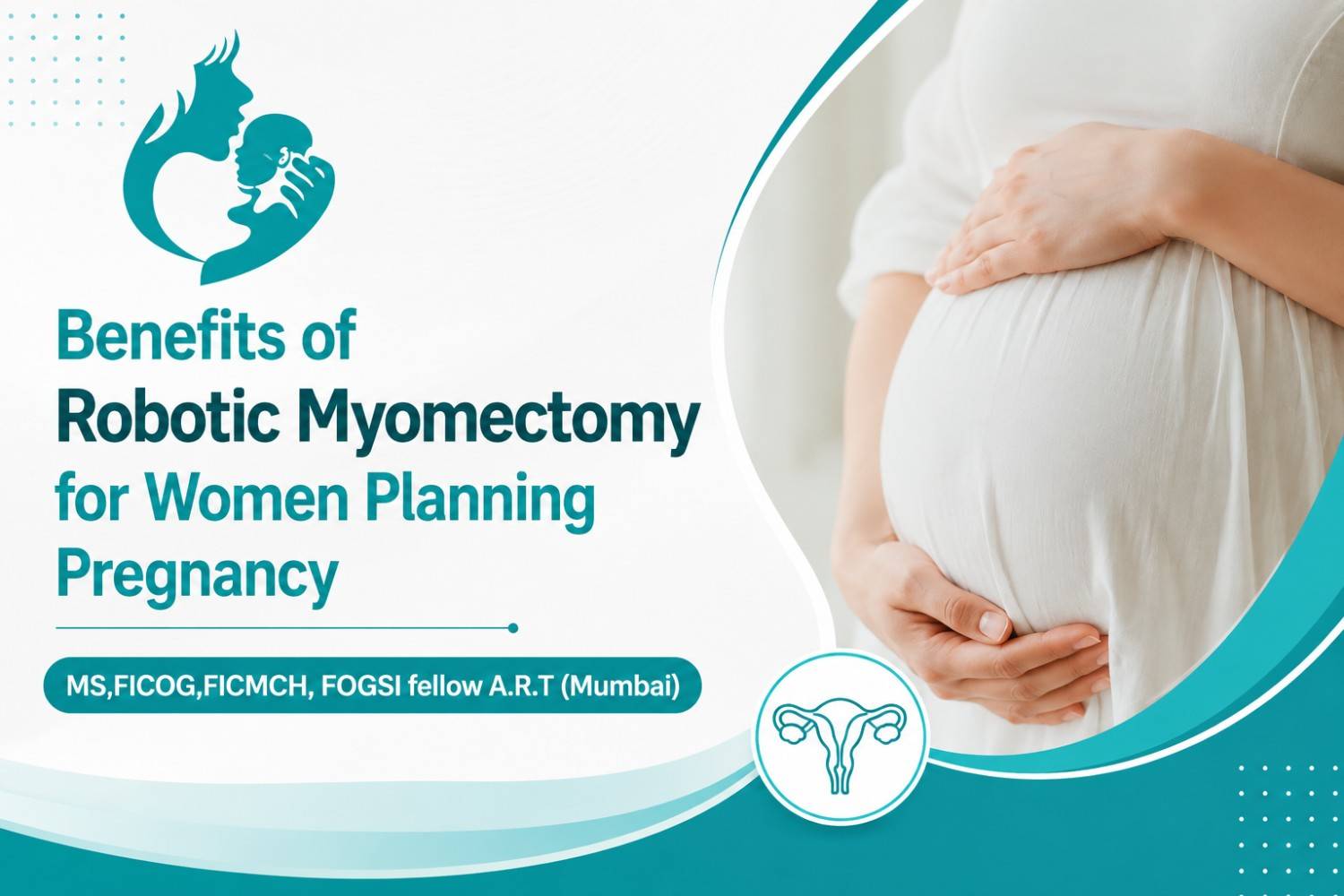
Common Orthopedic Problems in Pregnancy

Common Orthopedic Problems in Pregnancy

A woman experiences a lot of physical and emotional changes during pregnancy. To cope with them, it is important to consult the Best Gynecologist in Greater Kailash.

If we talk about physical changes in women during pregnancy, then orthopedic problems are common, and this is what we will be discussing here in the post.

Orthopedic Issues During Pregnancy

Lower Back Pain

Back pain is among the commonest orthopedic issues that a pregnant woman experiences. This is often because of the extra body weight. As a result of the growth and development of the fetus, the expecting mother gains weight. This puts pressure on the back muscles. Sometimes, the pain is severe. In such situations, having a strong back is important. So, maintain a strong back by doing exercises under the guidance of a skilled and experienced physiotherapist.

Osteitis Pubis

Another orthopedic condition that women may experience during pregnancy is osteitis pubis. It is an inflammatory condition that affects pubis symphysis. Bones at the front of the pelvis are called the pubic symphysis. Inflammation at this junction occurs because of the added weight and the position of the developing fetus. Pain associated with the condition can improve with the application of ice and heat and non-steroidal anti-inflammatory drugs (NSAIDs).

Heel Pain

Added body weight during pregnancy also causes heel pain. However, heel pain may occur because of plantar fasciitis. This is a condition characterized by the inflammation of a thick band of tissue present at the bottom of the foot. This band is called the plantar fascia and it connects the heel bone to the toes. Stabbing pain is what one experiences because of the condition which occurs during pregnancy. It is intense in the morning and the latter stages of pregnancy.

The application of ice and the use of shoe inserts may help improve the condition. In severe cases, the healthcare service provider may give cortisone shots.

Transient Osteoporosis of the Hip

This often occurs during late-term pregnancy. Transient hip osteoporosis is characterized by temporary bone loss around the hip joint. This makes the hip joint weak. The reason for the same is unknown but hormonal changes are believed to cause the same. Besides this, weight-bearing stress also contributes to the condition.

Knee Pain

Knee pain is very common in pregnancy and the reason for the same is simple i.e., added weight of carrying the baby. This is why rest is advised during pregnancy as it helps cope with knee pain by relieving pressure. This is especially true in the later stages of pregnancy as the fetus grows heavy.

To manage these conditions timely and ensure a healthy & happy pregnancy, consult an experienced doctor. Dr. Ruchi Tandon is an experienced gynecologist in South Delhi and also provides the best fibroids treatment in Greater Kailash.

Cervical infection in women

Cervicitis

Cervicitis is an inflammation of the cervix (the end of the uterus). Cervicitis often does not cause symptoms, but if they do happen, they may include abnormal vaginal discharge, painful intercourse, or vulvar or vaginal irritation. Antibiotics successfully treat cervicitis in most cases. For other cases, laser therapy or surgery may be required.

What is cervicitis?

Cervicitis is an inflammation of the cervix (the end of the uterus). This inflammation may be acute or chronic in nature. Cervicitis might not have symptoms, or a woman with cervicitis may have abnormal vaginal discharge.

Who gets cervicitis?

Cervicitis is very common. It has been projected that more than half of all adult women will have cervicitis at some point. Women who engage in high-risk sexual behaviors and have multiple partners are at a higher risk for cervicitis. Women who have had sexually transmitted diseases, or who have partners who have had sexually transmitted diseases, are also at a higher risk for cervicitis. Sexual practices are far from the only cause of the condition, however.

What are the causes of cervicitis?

There are many causes of cervicitis. Acute cervicitis is usually caused by an infection. Chronic cervicitis is more likely from a noninfectious cause.
Infections can result from the introduction of staphylococcus or streptococcus bacteria into the uterus. The sexually transmitted diseases chlamydia and gonorrhea are also significant causes of cervicitis. It has been estimated as much as 40 percent of cases of cervicitis are related to chlamydia. Herpes simplex virus, trichomonas and mycoplasma genitalium are also sometimes a cause.

Noninfectious causes of cervicitis include exposure to chemicals or

mechanical irritation. These include:

• Chemical irritation from spermicides or douches or the latex used in condoms
• Reaction to diaphragms, cervical caps, tampons or pessaries inserted
• Radiation therapy or systemic inflammatory diseases

What are the symptoms of cervicitis?

Symptoms of cervicitis can include:

• Abnormal vaginal discharge that may be yellow, white, or gray and have an unpleasant odor. This may persist for a long period of time.
• Sexual intercourse may become painful.
• Vulvar or vaginal irritation.

Cervicitis often does not have any symptoms, however. This is one reason why women should have regular gynecological appointments. It is especially important to seek an exam if you notice any of the above symptoms and/or have had chlamydia or another sexually transmitted disease in the past.

How is cervicitis diagnosed?

If you have any of the above symptoms, or if you have engaged in high-risk sexual practices, you should see your Gynecologist for a full pelvic exam. At this exam, your Gynecologist may perform tests including a Pap smear. In this test, a swab is used to collect cells from the cervix and uterus to look for abnormalities.

Your Gynecologist will also take a sample of any vaginal discharge to test for bacteria, such as bacterial vaginosis under a microscope. Tests for the sexually transmitted diseases gonorrhea and chlamydia will also be done.

Your Gynecologist also perform a full manual pelvic exam, looking for:

• Any redness on the cervix
• Discharge
• Blood on the vagina or cervix
• Inflammation of the vaginal walls
You will lay down on an examination table with your feet in stirrups for these tests.

How is cervicitis treated?

If your Gynecologist determines that you have cervicitis, you will usually be prescribed an antibiotic to treat any bacterial infections that may be causing the condition. Women under age 25 or who engage in high-risk behaviors may be treated with antibiotics even if bacteria are not detected in the diagnosis. Some bacteria can be hard to detect but may still be present. You should abstain from intercourse until treatment is done.

Antibiotics successfully treat cervicitis in most cases. If cervicitis is not successfully treated by antibiotics, laser therapy or surgery may be required. Your Gynecologist can best determine the treatment for your cervicitis based on your age, habits, diagnostic tests, and the length of the condition.

Dr. Ruchi Tandon is a reputed Gynecologist practicing in leading hospitals in South Delhi namely Max and Apollo hospitals with over 14 years of experience in handling all kinds of Gynecological conditions including cervical and vaginal infections .

COVID vaccine in women healthcare

Who should be vaccinated first?

As with all COVID-19 vaccines, health workers at high risk of exposure and older people should be prioritized for vaccination.

As more vaccine becomes available, additional priority groups should be vaccinated, with attention to people disproportionately affected by COVID-19 or who face health inequities.

Who else can take the vaccine?

The vaccine is safe and effective in people with known medical conditions associated with increased risk of severe disease, such as hypertension, diabetes, asthma, pulmonary, liver or kidney disease, as well as chronic infections that are stable and controlled.

Although further studies are required for immunocompromised persons, people in this category who are part of a group recommended for vaccination may be vaccinated after receiving information and counselling.

Persons living with HIV are at higher risk of severe COVID-19 disease. Known HIV-positive vaccine recipients should be provided with information and counselling.

Vaccination can be offered to people who have had COVID-19 in the past. But individuals may wish to defer their own COVID-19 vaccination for up to six months from the time of SARS-CoV-2 infection.

The vaccine can be offered to a breastfeeding woman who is part of a group recommended for vaccination (e.g. health workers); discontinuing breastfeeding after vaccination is currently not recommended.

Should pregnant women be vaccinated?

While pregnancy puts women at higher risk of severe COVID-19, very little data are available to assess vaccine safety in pregnancy.

Nevertheless, based on what we know about this kind of vaccine, we don’t have any specific reason to believe there will be specific risks that would outweigh the benefits of vaccination for pregnant women.

For this reason, those pregnant women at high risk of exposure to SARS-CoV-2 (e.g. health workers) or who have comorbidities which add to their risk of severe disease, may be vaccinated in consultation with their health care provider.

Who should not take the vaccine?

Individuals with a history of severe allergic reaction to any component of the vaccine should not take this or any other mRNA vaccine.

While vaccination is recommended for older persons due to the high risk of severe COVID-19 and death, very frail older persons with an anticipated life expectancy of less than 3 months should be individually assessed.

The vaccine should not be administered to persons younger than 18 years of age pending the results of further studies.

What’s the recommended dosage?

SAGE recommends the use of the Moderna mRNA-1273 vaccine at a schedule of two doses (100 µg, 0.5 ml each) 28 days apart. If necessary, the interval between the doses may be extended to 42 days.

Compliance with the full schedule is recommended and the same product should be used for both doses.

Is it safe?

While this vaccine has yet to be approved by WHO for an Emergency Use Listing, it has undergone review by the European Medicines Agency (EMA) and consequently meets WHO’s criteria for SAGE consideration.

The EMA has thoroughly assessed the data on the quality, safety and efficacy of the Moderna COVID-19 vaccine and authorized its use across the European Union.

SAGE recommends that all vaccinees be observed for at least 15 minutes after vaccination. Those who experience an immediate severe allergic reaction to the first dose should not receive additional doses.

Longer-term safety assessment involves continued follow up of clinical trial participants, as well as specific studies and continued surveillance of secondary effects or adverse events of those being vaccinated in the roll out.
The Global Advisory Committee on Vaccine Safety, a group of experts that provides independent and authoritative guidance to the WHO on the topic of safe vaccine use, receives and assesses reports of suspected safety events of potentially international impact.

How efficacious is the vaccine?

The Moderna vaccine has been shown to have an efficacy of approximately 92 per cent in protecting against COVID-19, starting 14 days after the first dose.

Does it work against new variants?

Based on the evidence so far, the new variants of SARS-CoV-2, including the B.1.1.7 and the 501Y.V2, do not alter the effectiveness of the Moderna mRNA vaccine. The monitoring, collection and analysis of data on new variants and their impact on the effectiveness of COVID-19 diagnostics, treatments and vaccines continues.

Dr. Ruchi Tandon is a reputed Gynecologist practicing in leading hospitals in South Delhi namely Max and Apollo hospitals with over 14 years of experience in handling all kinds of Gynecological conditions including high risk Pregnancy.

ecurrent miscarriage or recurrent Pregnancy loss

Spontaneous pregnancy loss is a surprisingly common occurrence, with approximately 15% of all clinically recognized pregnancies resulting in pregnancy failure. Recurrent pregnancy loss (RPL) has been inconsistently defined. When defined as 3 consecutive pregnancy losses prior to 20 weeks from the last menstrual period, it affects approximately 1% to 2% of women. This review highlights the current understanding of the various etiologies implicated in RPL, including factors known to be causative, as well as those implicated as possible causative agents. The appropriate diagnostic evaluation, therapy, and prognosis are also addressed.

Spontaneous pregnancy loss is a surprisingly common occurrence. Whereas approximately 15% of all clinically recognized pregnancies result in spontaneous loss, there are many more pregnancies that fail prior to being clinically recognized. Only 30% of all conceptions result in a live birth.

Spontaneous pregnancy loss can be physically and emotionally taxing for couples, especially when faced with recurrent losses. Recurrent pregnancy loss (RPL), also referred to as recurrent miscarriage or habitual abortion, is historically defined as 3 consecutive pregnancy losses prior to 20 weeks from the last menstrual period. Based on the incidence of sporadic pregnancy loss, the incidence of recurrent pregnancy loss should be approximately 1 in 300 pregnancies. However, epidemiologic studies have revealed that 1% to 2% of women experience recurrent pregnancy loss.

Defining RPL as a clinical entity requiring diagnostic testing and therapeutic intervention rests on knowledge of the elevation of risk for subsequent fetal loss and the probability of finding a treatable etiology for the disorder. Although no reliable published data have estimated the probability of finding an etiology for RPL in a population with 2 versus 3 or more miscarriages, the best available data suggest that the risk of miscarriage in subsequent pregnancies is 30% after 2 losses, compared with 33% after 3 losses among patients without a history of a live birth. This strongly suggests a role for evaluation after just 2 losses in patients with no prior live births. An earlier evaluation may be further indicated if fetal cardiac activity was identified prior to a loss, the woman is older than 35 years, or the couple has had difficulty in conceiving.

The high baseline rate of spontaneous isolated and recurrent pregnancy losses in the general population, the lack of consistent definition for RPL, limited access to tissues allowing study of the disorder, and the remarkably good prognosis for live birth among patients with RPL combine to frustrate aims at diagnostic and therapeutic recommendations. At present, there exist a small number of accepted etiologies for RPL.These include parental chromosomal abnormalities, untreated hypothyroidism, uncontrolled diabetes mellitus, certain uterine anatomic abnormalities, and antiphospholipid antibody syndrome (APS). Other probable or possible etiologies include additional endocrine disorders, heritable and/or acquired thrombophilias, immunologic abnormalities, infections, and environmental factors. After evaluation for these causes , approximately half of all cases will remain unexplained.

Genetic Etiologies

Approximately 2% to 4% of RPL is associated with a parental balanced structural chromosome rearrangement, most commonly balanced reciprocal or Robertsonian translocations. Additional structural abnormalities associated with RPL include chromosomal inversions, insertions, and mosaicism. Single gene defects, such as those associated with cystic fibrosis or sickle cell anemia, are seldom associated with RPL.

Appropriate evaluation of RPL should include parental karyotyping. Genetic counseling is indicated in all cases of RPL associated with parental chromosomal abnormalities. Depending on the particular diagnosis, directed therapy may include in vitro fertilization with preimplantation genetic diagnosis. The use of donor gametes may be suggested in cases involving genetic anomalies that always result in embryonic aneuploidy (ie, Robertsonian translocations involving homologous chromosomes).

Anatomic Etiologies

Anatomic abnormalities account for 10% to 15% of cases of RPL and are generally thought to cause miscarriage by interrupting the vasculature of the endometrium, prompting abnormal and inadequate placentation. Thus, those abnormalities that might interrupt the vascular supply of the endometrium are thought to be potential causes of RPL. These include congenital uterine anomalies, intrauterine adhesions, and uterine fibroids or polyps. Although more readily associated with second trimester losses or preterm labor, congenital uterine anomalies also play a part in RPL.

The uterine septum is the congenital uterine anomaly most closely linked to RPL, with as much as a 76% risk of spontaneous pregnancy loss among affected patients. Other Müllerian anomalies, including unicornuate, didelphic, and bicornuate uteri have been associated with smaller increases in the risk for RPL. The role of the arcuate uterus in causing RPL is unclear. The presence of intrauterine adhesions, sometimes associated with Asherman syndrome, may significantly impact placentation and result in early pregnancy loss.

Intramural fibroids larger than 5 cm, as well as submucosal fibroids of any size, can cause RPL. Although congenital anomalies caused by prenatal exposure to diethylstilbestrol are clearly linked to RPL, this is becoming less clinically relevant as most affected patients move beyond their reproductive years.

Diagnostic evaluation for uterine anatomic anomalies should include office hysteroscopy or hysterosalpingography (HSG). Hysteroscopic resection of intrauterine adhesions and intrauterine septa are indicated if these abnormalities are identified. Patients undergoing successful hysteroscopic septum resection seem to enjoy near normal pregnancy outcomes, with term delivery rates of approximately 75% and live birth rates approximating 85%.Myomectomy should be considered in cases of submucosal fibroids or any type fibroids larger than 5 cm. Resection has been shown to significantly improve live birth rates from 57% to 93%.Myomectomy can be performed via open laparotomy, laparoscopy, or hysteroscopy.

Dr. Ruchi Tandon is a reputed Gynecologist practicing in leading hospitals in South Delhi namely Max and Apollo hospitals with over 14 years of experience in handling all kinds of Gynecological conditions including recurrent abortions.

Abnormalities in uterus or reproductive organs

An understanding of congenital anomalies as they are encountered in clinical practice is greatly enhanced by not only a knowledge of normal embryology and the mechanism of formation of normal infants, but also an insight into the processes that result in the development of anomalies. An awareness of malformations and a systematic examination and appraisal of every neonate will greatly increase the number of such anomalies found. In some instances, e.g., congenital adrenal hyperplasia, imperforate anus, diaphragmatic hernia, and esophageal atresia, early detection and prompt intervention may be lifesaving. In adults, amenorrhea is an important clue and may suggest an imperforate hymen, vaginal septum or absence of the uterus.

The finding of one anomaly should stimulate a careful gynecologist to carry out a complete study to detect renal and ureteral anomalies, particularly the solitary pelvic kidney which might be removed as a “pelvic mass”. Many anomalies occur infrequently so that only physicians in large medical centers may see them frequently enough to be aware of the possible anomalies and their causation, prognosis, and, in some cases, correction. The identification and interpretation of such abnormalities constitute a real challenge to the clinician. A knowledge of the problems and pitfalls in the management of these defects will benefit both the obstetrician and the gynecologic surgeon.

Causes of abnormalities

The causes of congenital malformations or abnormalities present at birth may be either environmental or genetic (chromosomal abnormalities). It is not always easy to separate the two factors; both may be at work in the same embryo or fetus. Rapidly growing embryonic organs are the most sensitive to environmental influences.

Millen has classified the mechanisms of anomaly production as follows:

1. Developmental arrest—cessation of development before completion
2. Agenesis or aplasia—failure of normal development
3. Hyperplasia or local overgrowth
4. Aberrant development
5. Failure of normal resorption (either too much or too little) or resorption in the wrong locations
6. Secondary degeneration of normally developed structures

Millen also emphasizes “that the period when environmental agents may affect the development of an embryo is very short, being nearly over by the end of the eighth week of pregnancy”. Organogenesis occurs from day 13 to day 60; teratogenic (G. teras, monster) agents are most dangerous during this period. There is a time relationship between specific organ systems and sensitivity to environmental factors as well as a relationship between specific teratogens and specific organ systems. Examples are rubella infections occurring in the first trimester, with a high incidence of cataracts, deafness, and cardiac malformations, and use of thalidomide, with varied malformations of arms and legs.

Nutgen has evaluated in detail the mechanisms of action of various environmental teratogenic factors. These include the following:

1. Ionizing radiation
2. Vital disease and related infections
3. Chemical factors
4. Immunologic disturbances
5. Hormones
6. Nutritional factors

Ionizing radiation is probably one of the best known damaging factors. Infections such as rubella virus, cytomegalovirus, and Toxoplasma gondii can cause severe damage to the eyes and central nervous system. Chemicals include aminopterin (causing skeletal defects and nervous system damage), methotrexate, and thalidomide. Immunologic disturbances include Rhesus incompatibilities. Hormone damage is particularly interesting: the administration of exogenous testosterone, synthetic progestogens, and similar preparations can cause iatrogenic deformities of the female genitalia.

Pathologic hyperandrogenemia, as seen in luteomas of pregnancy, can result in virilization in the female newborn. Environmental factors, such as exposure to diesel fumes, have also been associated with virilization due to inhibition of aromatase and accumulation of excess testosterone. Nutritional factors apparently have little direct teratogenic effect on the fetus.

Uterine abnormalities

The most frequent uterine anomalies (Fig. 3) are those resulting from varying degrees of failure of fusion of the müllerian ducts. This variability makes classification difficult and determination of the true incidence uncertain. Many of these malformations are detected by radiologic or sonographic studies. Estimates of incidence vary from 0.13% to 4.0%. The incidence of müllerian anomalies in patients with infertility has been reported to be as high as 6.3%.Pregnancy occurs in many women despite these anomalies. The complication rates with pregnancy are considerably increased; complications include abortion, prematurity, postpartum hemorrhage, retained placenta, and breech presentation. Not surprisingly, the rate of cesarean delivery is markedly higher.

Uterus duplex, or the bicornuate uterus, is the most frequent uterine anomaly. The unicollis type in which there is a single cervix with a septum that does not reach the cervix is the most frequent type, occurring in over one third of all patients with uterine anomalies.

Uterus duplex bicollis, in which two cervices are present, is less frequent. Obstetric complications are frequent, but live births do occur.

Uterus didelphys, with completely separate uterine cavities, is also frequent. The cervices are externally united and the uterine fundi are externally separate. In most patients the vagina is septate, causing a double vagina. The halves of such a uterus are often of different sizes. If there is an asymmetric vaginal septum which occludes one vagina, mucocolpos or hematocolpos may result. Communicating uteri, involving an incomplete uterine septum with part of the fetus in each uterine cavity, occasionally occur.

Uterus septus is an essentially normal uterus with a septum reaching to the cervix.

Uterus subseptus involves a partial septum that does not reach the cervix. Twins apparently occur approximately three times more often in women with this condition than in women with normal uteri; the cause is not clear.
Uterus arcuatus is a normal uterus without a septum. The fundus, however, is notched or flattened. There is usually no interference with normal pregnancy.
Uterus unicornis is a uterus with a single horn. A normal vagina and a single normal tube are usually present. The other half of the uterus is usually absent or rudimentary. In most patients the kidney is missing on the side of the missing uterus. Successful pregnancy can occur.

Separate hemiuteri with separate vaginas is a rare condition that is usually associated with duplications of urethra and bladder or of the colon and anus. Pregnancy in each of the two hemiuteri in the same woman at different times has been reported.

These anomalies result from failure of fusion of the paired müllerian ducts, but in some instances there is a true duplication of the ducts on one or both sides. Such duplications result from splitting of the müllerian duct during the seventh week of development. Accessory tubes or ovaries may be present.

Semmens extensively reviewed the literature on genital tract anomalies and evaluated 56 personal patients as well as 500 cases from the literature. He employed a simplified classification, based entirely on the functional capacity of the uterine cavity, which divided genital tract anomalies into two groups: group I—hemiuterus of single müllerian origin, paired or otherwise, and group II—uterus of dual müllerian origin, associated with varying degrees of absorption. He concluded that if the entire functional component has been derived from a single müllerian duct and its vaginal outlet is a cervical canal of similar origin its capacity is smaller than that of the uterus resulting from fusion of bilateral ducts.

By contrast, if the uterus is derived from two müllerian ducts, its capacity is larger. Semmens concluded that the space available for the developing fetus as well as the variations in uterine circulation had a direct effect on the length of gestation, onset and behavior of labor, and overall fetal salvage. The diagnosis of uterine anomalies during pregnancy is always difficult. The most important factor is awareness of their existence and frequency and of the problems that frequently develop. Findings such as a floating head at term without apparent cause, notching and broadening of the uterine fundus, abnormal lie, recurrent breech presentations, prolonged third stage of labor, and trapped or retained placenta indicate the possibility of an anomaly. In abnormal uteri, triangular spasm and cornual pocketing of the placenta may occur and necessitate manual removal of the placenta.

The administration of oxytocin under these circumstances usually increases the degree of pocketing in an abnormal uterus. Among antepartum patients, pyelitis (often associated with a urologic anomaly), passage of a decidual cast, hemorrhage, and premature rupture of the membranes are the most common complications. Intrapartum complications include septal dystocia, incarcerated nongravid horn, uterine inertia, and retained placenta. Fetal dystocia is considerably increased. Postpartum complications are few and include hemorrhage, retained placenta, and failure of uterine involution. Most complications of uterine anomalies in the pregnant woman can be anticipated and managed successfully if the clinician is alert to the possibility of anomalies and if the nature of the anomaly is known.

The finding of an asymmetrically located cervix in the vaginal fornix, an excessively large cervix whether or not a septum is present, or a duplicated cervix suggests uterine abnormality. Abnormal configurations of the fundus of the uterus in the third trimester should suggest abnormalities to the clinician, as should abnormal presentations and failure of the presenting part to engage without apparent reason. An obstetric history of successive abortions, recurrent breech presentations, or recurrent pyelitis should alert the obstetrician to the possibility of anomalies and, in nonpregnant women, to the need for hysterosalpingogram, saline sonography, magnetic resonance imaging, and renal imaging.

Dr. Ruchi Tandon is a reputed Gynecologist practicing in leading hospitals in South Delhi namely Max and Apollo hospitals with over 14 years of experience in handling all kinds of Gynecological conditions including uterine abnormalities.

Diagnostic Laparoscopy for infertility

Laparoscopy overview

• Laparoscopy for infertility is a minimally invasive surgical procedure in which a telescope-like instrument (laparoscope) with light and small camera allows the surgeon to examine the pelvic anatomy for causes of female infertility.
• Laparoscopy can be diagnostic, assessing for causes of infertility in the uterus, fallopian tubes and ovaries.
• An operative laparoscopy involves surgical treatment for problems found during a diagnostic laparoscopy procedure, utilizing small surgical tools passed through the laparoscope.
• Laparoscopic surgery can also be used to remove scar tissue or endometriosis.

When is laparoscopy used for treating infertility?

Laparoscopy for infertility is generally only performed after other fertility tests have not resulted in a conclusive diagnosis. For this reason, laparoscopy is often performed on women with unexplained infertility.

Laparoscopy also allows for biopsy of suspect growths and cysts that may be hampering fertility. Laparoscopy may be recommended for women experiencing pelvic pain, which is a potential symptom of endometriosis. Laparoscopy can also remove scar tissue that can be a cause of pelvic or abdominal pain.

Why It’s Needed

Some causes of infertility can only be diagnosed through laparoscopy.

(Endometriosis, for example.) Laparoscopy allows your doctor to not only see what’s inside your abdomen but also biopsy suspicious growths or cysts.

Also, laparoscopic surgery can treat some causes of infertility, allowing you a better chance at getting pregnant either naturally or with fertility treatments.

How Is It Done?

Laparoscopy is performed in a hospital under general anesthesia. While it is sometimes possible to conduct a diagnostic laparoscopy in a fertility clinic office, this is not recommended. In the office setting, if something is found during the procedure, you will need to have the procedure again in a hospital setting for the repair.

Your doctor will give you instructions on how to prepare for surgery beforehand. You will probably be told not to eat or drink for 8 or more hours before your scheduled surgery, and you may be instructed to take antibiotics.

When you get to the hospital, you’ll receive an IV, through which fluids and medication to help you relax will be delivered. The anesthesiologist will place a mask over your face, and after breathing a sweet-smelling gas for a few minutes, you’ll fall asleep.

Once the anesthesia has taken effect, the doctor will make a small cut around your belly button. Through this cut, a needle will be used to fill your abdomen with carbon dioxide gas. This provides room for your doctor to see the organs and move the surgical instruments.

Once your abdomen is filled with gas, the surgeon will then place the laparoscope through the cut to look around at your pelvic organs. The surgeon may also biopsy tissue for testing.

The surgeon will visually evaluate the pelvic organs and the surrounding abdominal organs. He or she will look for the presence of cysts, fibroids, scar tissue or adhesions, and endometrial growths. He or she will also look at the shape, color, and size of the reproductive organs.

A dye may be injected through the cervix, so the surgeon can evaluate if the fallopian tubes are open.

Even if no signs of endometriosis or other problems are found, the surgeon may remove a sample of tissue to be tested. Sometimes, very mild endometriosis is microscopic and cannot be seen by the naked eye with the laparoscopic camera.

If an ectopic pregnancy is suspected, the surgeon will evaluate the fallopian tubes for abnormal pregnancy.

How Will It Feel?

During laparoscopic surgery, you’ll be under the effects of general anesthesia, so you should not feel any pain, nor remember the procedure.

When you wake up, you may have a sore throat. This is caused by the tube placed down your throat to help you breathe during surgery. (This tube is removed before you wake-up).

It’s normal for the area around the cuts to feel sore, and your abdomen may feel tender, especially if your doctor removes a lot of scar tissue. You may feel bloated from the carbon dioxide gas, and you may experience sharp pains in your shoulder. This should go away in a few days.

Dr. Ruchi Tandon is a reputed Gynecologist practicing in leading hospitals in South Delhi namely Max and Apollo hospitals with over 14 years of experience in handling all kinds of Gynecological conditions including infertility.

Blocked fallopian tubes

Blocked fallopian tubes are one possible cause of female infertility. There are usually no symptoms, but there are some risk factors that can increase the chance of developing the condition. The medical term for a blocked fallopian tube is tubal occlusion.

The fallopian tubes are muscular tubes that are lined with delicate hair-like structures. These “hairs” work in both directions; helping an egg to travel from the ovaries down to the womb (uterus) and helping sperm travel up from the womb.

Each fallopian tube ends in fimbriae, which are finger-like structures. The fimbriae catch and guide an egg when the ovary releases it.

The fallopian tubes play an important role in conception because they are where most eggs are fertilized.

If any part of the fallopian tube is damaged, for example by surgery or an infection, they can become blocked by scar tissue.

Symptoms

Blocked fallopian tubes do not often present symptoms other than difficulty conceiving. Doctors typically class this as having tried to conceive for 1 year without success.

A blocked fallopian tube may cause some women to experience symptoms such as pain in the pelvis or belly. This pain might happen regularly, such as around the time of their period, or be constant.

Sometimes, a blockage in a fallopian tube can cause a fertilized egg to get stuck. This is known as an ectopic pregnancy.

An ectopic pregnancy may not always cause symptoms and is usually detected during a scan. However, some woman may experience signs of pregnancy, such as stomach pain on one side of the body, or vaginal bleeding. Any woman who suspects she has an ectopic pregnancy should seek immediate medical attention.

Causes

Fallopian tubes can become blocked for a range of reasons, which include:
• a history of pelvic infection
• a previous burst appendix
• having had a sexually transmitted disease, such as gonorrhea or chlamydia
• endometriosis, a condition that causes the lining of the womb to grow outside of the uterus
• history of abdominal surgery
• hydrosalpinx, which is swelling and fluid at the end of a fallopian tube Effects on fertility

All of these conditions can affect the fallopian tubes directly or this area of the body. In most cases, these conditions or procedures create scar tissue that can block the tubes.

The female reproductive system is made up of the ovaries, uterus, and fallopian tubes.

If a medical problem has affected any of these three areas, it may make getting pregnant more difficult.

Each of the two ovaries is connected to the uterus by a fallopian tube. The ovaries store eggs and release them randomly, with one ovary releasing an egg each month.

For example, the right ovary might release an egg for 3 months in a row, and then the left ovary might release an egg the following month.

If one fallopian tube is blocked, it may still be possible for an egg to be fertilized. If both are blocked, this is less likely.

Diagnosis

Blocked fallopian tubes can be difficult to identify. The tubes can open and close, so it is not always easy to tell if they are blocked or just closed.

There are three key tests to diagnose blocked fallopian tubes:

• An X-ray test, known as a hysterosalpingogram or HSG. A doctor injects a harmless dye into the womb, which should flow into the fallopian tubes. The stain is visible on an X-ray. If the fluid does not flow into the fallopian tubes, they may have a blockage.
• An ultrasound test, known as a sonohysterogram. This is very similar to the HSG test but uses sound waves to build up a picture of the fallopian tubes.
• Keyhole surgery, known as a laparoscopy. A surgeon makes a small cut in the body and inserts a tiny camera to take pictures of the fallopian tubes from inside.

A laparoscopy is the most accurate test for blocked tubes. However, doctors may not recommend this test as an early diagnosis because it is invasive and cannot treat the issue.

A doctor may be able to suggest a possible diagnosis based on medical history. For example, a woman may have had a burst appendix in the past. If the woman has had difficulty conceiving, this could suggest blocked fallopian tubes as a likely cause.

Dr. Ruchi Tandon is a reputed Gynecologist practicing in leading hospitals in South Delhi namely Max and Apollo hospitals with over 14 years of experience in handling all kinds of Gynecological conditions including high risk Pregnancies and blocked fallopian tubes.

Vaccination in pregnancy

Immunization during pregnancy is a simple and effective way to protect the mother and child from certain infections. The immunological changes occur during pregnancy which may be responsible for the susceptibility of certain infectious diseases that increases the risk of more serious outcomes. Vaccination of pregnant women can protect to mother against vaccine-preventable infections, and in so doing potentially protect the fetus. Immunization during pregnancy can also directly protect the fetus and infant via transferred of antibodies from the mother to the fetus.

This is why vaccinations during pregnancy are so important. Vaccination during pregnancy is a cost-effective strategy to improve pregnancy outcomes in India. Globally, no scientific study exist which shows the risk of fetus after vaccination of pregnant women with inactivated vaccines or bacterial vaccines or toxoids. Even live vaccines causing risk to fetus is theoretical. Vaccination with inactivated virus, bacterial or toxoid in pregnancy is risk to a developing fetus during pregnancy is theoretical. But definitely the live vaccine poses a theoretical risk to a developing fetus. Therefore, all live vaccines should be avoided during pregnancy. The developing country like India where the people can’t afford these vaccines, the government should be included these vaccines in routine immunization program.

Maternal immunization provides important health benefits to both pregnant women and to their fetus. Vaccine-preventable diseases cause significant morbidity and mortality among maternal, neonatal, and young infant. Some infections are so serious even they can waste pregnancy, harm her baby during pregnancy or after delivery. These complications can be protected with vaccination. This is why vaccinations are so important for pregnant mothers. Vaccines strengthen the immune systems of body that can fight off serious infectious diseases.

A vaccine can help in protection of the mother’s body from infections and this immunity passes to her baby during pregnancy. This immunity keeps the child safe during the first few months of life until baby gets his own vaccination. Vaccination also protects mothers from getting a serious disease that could affect future pregnancies. Fetus getting any risk after vaccination of the mother during pregnancy primarily is theoretical. Globally, no scientific study exist which shows the risk of fetus after vaccination of pregnant women with inactivated vaccines or bacterial vaccines or toxoids.

Even live vaccines causing risk to fetus is theoretical. Benefits of vaccinating pregnant women usually outweigh potential risks when the likelihood of disease exposure is high, when infection would pose a risk to the mother or fetus, and when the vaccine is unlikely to cause harm. Not all vaccinations are safe during pregnancy but some of inactivated vaccines are considered safe which can be give to pregnant women who might be at risk of infection.

Tetanus vaccine

Tetanus can cause severe morbidity in the mother and mortality in the neonate. Tetanus is a life-threatening bacterial disease that is caused by the toxin of a bacterium called Clostridium tetani which is often found in soil. Tetanus bacteria enter the body through an open wound. It could well be a tiny prick or scratch on the skin, although Tetanus infection is more common when there is a deep puncture wound such as a bite, cut, burn or an ulcer.

Tetanus affects a person’s nervous system and can be fatal if left untreated. Neonatal tetanus usually occurs in newborns through infection of the unhealed umbilical stump, especially when the stump is cut with a non-sterile instrument. Tetaus is prevented only through vaccination. The tetanus vaccine contains noninfectious toxoids. Tetanus toxoids appear safe during pregnancy and are administered in many countries of the world to prevent neonatal tetanus.

The World Health Organization (WHO) reported that neonatal tetanus kills over 200,000 newborns each year; almost all these deaths occur in developing countries while it is very rare in developed nations.

To maximize the maternal antibody response and passive antibody transfer to the infant, the national immunization schedule in India recommends the 2 doses of tetanus toxoid (TT) for unknown immunization status of pregnant women i.e the first dose of tetanus toxoid should be administered as soon as pregnancy is detected, second dose of tetanus toxoid is administered after 4 weeks and if a mother received 2 TT doses in the last pregnancy and mother gets again pregnant with in 3 y than only one dose of TT is recommended and that dose is called booster dose. Some experts recommend that the second dose of the vaccine should be given 4 weeks prior to the expected date of delivery.

The WHO also recommends that a third vaccine be given 6 months after the second one to provide protection for at least 5 y After TT vaccination, the antibodies formed in mother are transferred to baby and protect baby for a few months after birth. TT vaccination also helps to prevent premature birth or delivery. The American Congress of Obstetricians and Gynecologists recommends use of tetanus immune globulin, as there is no evidence of any adverse effects to the fetus from the tetanus immunoglobin.

Influenza vaccine

Influenza can cause severe illness in pregnant women than non pregnant women. During pregnancy, there is a change in the immune system, heart and lungs which makes pregnant women more prone to severe illness as well as hospitalizations and even death from influenza. Influenza infection during pregnancy also has a greater chance for serious problems for their unborn babies, including premature labor and delivery.

Women in the second and third trimesters of pregnancy are at increased risk for hospitalization from influenza. Therefore, routine inactivated influenza vaccine is recommended for all women who will be pregnant (in any trimester) during influenza season. The available inactivated influenza vaccines for the control of seasonal influenza are safe and efficacious and have the potential to prevent significant morbidity and mortality in pregnant women.

Live attenuated influenza vaccine is available in form of an intranasal spray and that is not recommended for pregnant women. Maternal influenza immunization offers demonstrated disease prevention benefits for women and their newborns and is a critically important component during pregnancy. Pregnant women should be counseled about the benefits of the influenza vaccine for themselves and their unborn baby.

Dr.Ruchi Tandon is a reputed Gynecologist practicing in leading hospitals in South Delhi namely Max and Apollo hospitals with over 14 years of experience in handling all kinds of Gynecological conditions including high risk pregnancy .

Abnormal blood pressure in pregnancy

Blood pressure and pregnancy

During pregnancy, your body goes through numerous physical changes to accommodate fetal growth and development. Throughout these nine months, it’s ideal to have a normal blood pressure reading.

Your blood pressure is the force of your blood pushing against the walls of your arteries. Every time your heart beats, it pumps blood into the arteries, which then carry the blood to the rest of your body. The blood typically moves through the arteries at a certain rate. However, various factors can disrupt the normal rate at which blood flows through the vessels, causing an increase or decrease in pressure. Increased pressure in the arteries can result in an elevated blood pressure reading. Decreased pressure in the arteries can result in a low blood pressure reading.

Blood pressure is recorded as two types of numbers. The systolic number is the top number, which indicates the amount of pressure in the arteries when your heart beats. The diastolic number is the bottom number, which indicates the amount of pressure in the arteries between heartbeats. Your blood pressure naturally rises with each heartbeat and falls when the heart rests in between beats. However, the rapid changes your body goes through during pregnancy can greatly influence these numbers and cause a drastic change in blood pressure.

According to the American Heart Association (AHA), a normal blood pressure reading is 120/80 mm Hg and below. Readings below 90/60 mm Hg indicate low blood pressure, or hypotension. Readings above 140/90 mm Hg in pregnancy indicate high blood pressure, or hypertension. Hypertension is seen far more often in pregnancy than hypotension. About 10 percent of all pregnancies in the United States are complicated by high blood pressure issues.

An abnormal blood pressure during pregnancy is a cause for concern. Both you and your baby may be at an increased risk of health complications. However, you may be able to prevent problems by attending regular prenatal appointments so your doctor can monitor your blood pressure closely. You may also want to consider learning more about related conditions so you can help manage factors affecting your blood pressure.

How to detect abnormal blood pressure

The AHA defines abnormal blood pressure readings in non-pregnant adults as follows:

• Elevated blood pressure is a systolic number between 120 and 129 and a diastolic number lower than 80.
• In stage 1 hypertension, the systolic number is between 130 and 139 or the diastolic number is between 80 and 89.
• In stage 2 hypertension, the systolic number is 140 or higher or the diastolic number is 90 or higher.
• In a hypertensive crisis, the systolic number is higher than 180 and/or the diastolic number is higher than 120.

You may not always be able to tell whether your blood pressure is too high or too low. In fact, hypertension and hypotension may not cause noticeable symptoms. If you do experience symptoms, they may include the following:

Symptoms of hypertension

High blood pressure, or hypertension, in pregnancy is usually defined as 140/90 mm Hg or higher. It may cause:

• flushed skin
• swelling of the hands or feet
• headaches
• shortness of breath
• abdominal pain
• nausea
• vomiting
• changes in vision

Symptoms of hypotension

Low blood pressure, or hypotension, is usually defined as 90/60 mm Hg or less. It may cause:

• dizziness
• difficulty concentrating
• cold, clammy skin
• blurred vision
• rapid breathing
• depression
• sudden tiredness
• extreme fatigue

If you suspect you have symptoms of hypertension or hypotension, you should see your doctor right away to help prevent possible complications.

The symptoms of hypertension and hypotension aren’t always present. The best way to know if you have abnormal blood pressure is to take a blood pressure test. Blood pressure tests are often done at regular checkup appointments, and your doctor should perform them throughout your pregnancy.

While these tests are most commonly performed in a medical setting, they can also be done at home. Many local drug stores carry at-home blood pressure monitors that you can use to check your blood pressure. However, make sure to consult with your doctor before you try monitoring your blood pressure at home. Your doctor may have specific instructions regarding when and how often you should check your blood pressure.

Causes of abnormal blood pressure during pregnancy

The AHA estimates that 1 out of every 3 American adults has hypertension. In pregnancy, hypertension can be classified into two main categories: chronic hypertension and hypertension related to pregnancy. Chronic hypertension refers to high blood pressure that was present before pregnancy. You may also be diagnosed with this condition if you develop hypertension during the first 20 weeks of pregnancy. You may still have the condition after giving birth.

High blood pressure disorders related to pregnancy generally develop after the first 20 weeks of pregnancy. There are several types of disorders that range in severity. A 2016 review published in Integrated Blood Pressure Control Trusted Source suggests that age, obesity, and underlying health problems seem to contribute to these conditions. Though these conditions usually go away after you give birth, your risk of getting hypertension in the future is much higher if you develop any of them.

Hypotension, while much less common, can be directly related to pregnancy. Your circulatory system expands during pregnancy to accommodate your fetus. As circulation expands, you might experience a small drop in blood pressure. According to the AHA, this is most common during the first 24 weeks of pregnancy. Still, this amount is usually not significant enough to cause concern.

Hypotension may also be caused by:

• dehydration
• diabetes
• low blood sugar
• heart problems
• thyroid problems
• severe allergic reactions
• blood loss
• infection
• malnutrition, particularly a lack of folic acid, B vitamins, and vitamin D

Dr. Ruchi Tandon is a reputed Gynecologist practicing in leading hospitals in South Delhi namely Max and Apollo hospitals with over 14 years of experience in handling all kinds of Gynecological conditions and Pregnancies.

Urinary Tract infections in females

A urinary tract infection (UTI) is an infection from microbes. These are organisms that are too small to be seen without a microscope. Most UTIs are caused by bacteria, but some are caused by fungi and in rare cases by viruses. UTIs are among the most common infections in humans.

A UTI can happen anywhere in your urinary tract. Your urinary tract is made up of your kidneys, ureters, bladder, and urethra. Most UTIs only involve the urethra and bladder, in the lower tract. However, UTIs can involve the ureters and kidneys, in the upper tract. Although upper tract UTIs are more rare than lower tract UTIs, they’re also usually more severe.

UTI symptoms

Symptoms of a UTI depend on what part of the urinary tract is infected.

Lower tract UTIs affect the urethra and bladder. Symptoms of a lower tract UTI include:

• burning with urination
• increased frequency of urination without passing much urine
• increased urgency of urination
• bloody urine
• cloudy urine
• urine that looks like cola or tea
• urine that has a strong odor
• pelvic pain in women
• rectal pain in men

Upper tract UTIs affect the kidneys. These can be potentially life threatening if bacteria move from the infected kidney into the blood. This condition, called urosepsis, can cause dangerously low blood pressure, shock, and death.

Symptoms of an upper tract UTI include:

• pain and tenderness in the upper back and sides
• chills
• fever
• nausea
• vomiting

UTI treatment

Treatment of UTIs depends on the cause. Your doctor will be able to determine which organism is causing the infection from the test results used to confirm the diagnosis.

In most cases, the cause is bacteria. UTIs caused by bacteria are treated with antibiotics.

In some cases, viruses or fungi are the causes. Viral UTIs are treated with medications called antivirals. Often, the antiviral cidofovir is the choice to treat viral UTIs. Fungal UTIs are treated with medications called antifungals.

Antibiotics for a UTI

The form of antibiotic used to treat a bacterial UTI usually depends on what part of the tract is involved. Lower tract UTIs can usually be treated with oral antibiotics. Upper tract UTIs require intravenous antibiotics. These antibiotics are put directly into your veins.

Sometimes, bacteria develop resistance to antibiotics. To reduce your risk of antibiotic resistance, your doctor will likely put you on the shortest treatment course possible. Treatment typically lasts no more than 1 week.

Results from your urine culture can help your doctor select an antibiotic treatment that will work best against the type of bacteria that’s causing your infection.

Treatments other than antibiotics for bacterial UTIs are being examined. At some point, UTI treatment without antibiotics may be an option for bacterial UTIs by using cell chemistry to change the interaction between the body and the bacteria.

UTI diagnosis

If you suspect that you have a UTI based on your symptoms, contact your doctor. Your doctor will review your symptoms and perform a physical examination. To confirm a diagnosis of a UTI, your doctor will need to test your urine for microbes.

The urine sample that you give your doctor needs to be a “clean catch” sample. This means the urine sample is collected at the middle of your urinary stream, rather than at the beginning. This helps to avoid collecting the bacteria or yeast from your skin, which can contaminate the sample. Your doctor will explain to you how to get a clean catch.

When testing the sample, your doctor will look for a large number of white blood cells in your urine. This can indicate an infection. Your doctor will also do a urine culture to test for bacteria or fungi. The culture can help identify the cause of the infection. It can also help your doctor choose which treatment is right for you.

If a virus is suspected, special testing may need to be performed. Viruses are rare causes of UTIs but can be seen in people who have had organ transplants or who have other conditions that weaken their immune system.

Upper tract UTIs

If your doctor suspects that you have an upper tract UTI, they may also need to do a complete blood count (CBC) and blood cultures, in addition to the urine test. A blood culture can make certain that your infection hasn’t spread to your blood stream.

Recurrent UTIs

If you have recurrent UTIs, your doctor may also want to check for any abnormalities or obstructions in your urinary tract. Some tests for this include:

• An ultrasound, in which a device called a transducer is passed over your abdomen. The transducer uses ultrasound waves to create an image of your urinary tract organs that are displayed on a monitor.
• An intravenous pyelogram (IVP), which involves injecting a dye into your body that travels through your urinary tract and taking an X-ray of your abdomen. The dye highlights your urinary tract on the X-ray image.
• A cystoscopy, which uses a small camera that’s inserted through your urethra and up into your bladder to see inside your bladder. During a cystoscopy, your doctor may remove a small piece of bladder tissue and test it to rule out bladder inflammation or cancer as a cause of your symptoms.
• A computerized tomography (CT) scan to get more detailed images of your urinary system.

Causes and risk factors of a UTI

Anything that reduces your bladder emptying or irritates the urinary tract can lead to UTIs. There are also many factors that can put you at an increased risk of a getting a UTI. These factors include:

• age — older adults are more likely to get UTIs
• reduced mobility after surgery or prolonged bed rest
• kidney stones
• a previous UTI
• urinary tract obstructions or blockages, such as an enlarged prostate, kidney stones, and certain forms of cancer
• prolonged use of urinary catheters, which may make it easier for bacteria to get into your bladder
• diabetes, especially if poorly controlled, which may make it more likely for you to get a UTI
• pregnancy
• abnormally developed urinary structures from birth
• a weakened immune system

Dr. Ruchi Tandon is a reputed Gynecologist practicing in leading hospitals in South Delhi namely Max and Apollo hospitals with over 14 years of experience in handling all kinds of Gynecological conditions including urinary tract infections.