Women’s healthcare has witnessed remarkable progress over the last decade, and robotic-assisted surgery is one of the most significant advancements. From treating fibroids to performing complex gynecological procedures with greater precision, robotic technology is helping women recover faster and experience better outcomes. Today, many women seeking advanced treatment options prefer consulting the best gyno doctor in South Delhi for minimally invasive robotic procedures that offer improved comfort and safety.
Understanding Robotic Gynecologic Surgery
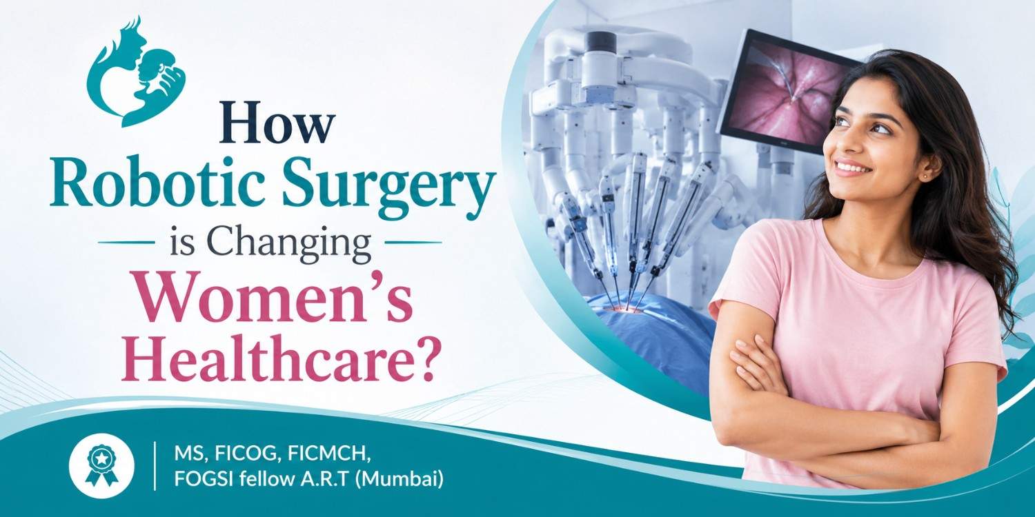
Robotic surgery is a minimally invasive surgical technique where surgeons use robotic arms controlled through a specialized console. The system provides a high-definition 3D view and allows highly precise movements during surgery. Unlike traditional open surgeries, robotic procedures require only small incisions, reducing trauma to surrounding tissues.
This modern approach is commonly used for treating:
- Uterine fibroids
- Endometriosis
- Ovarian cysts
- Gynecologic cancers
- Hysterectomy procedures
- Fertility-related conditions
The growing adoption of robotic surgery is transforming how gynecological treatments are performed while improving patient comfort.
Key Benefits of Robotic Surgery for Women
Smaller Incisions and Minimal Scarring
Traditional surgeries often involve large incisions that may take weeks to heal. Robotic-assisted procedures use tiny cuts, which means less visible scarring and reduced postoperative discomfort.
Faster Recovery Time
One of the biggest advantages of robotic surgery is quicker recovery. Most patients can resume daily activities sooner compared to conventional surgery. This is especially beneficial for working women and mothers managing busy schedules.
Greater Surgical Precision
Robotic systems enhance the surgeon’s control and accuracy. Delicate procedures involving reproductive organs can be performed with improved precision, reducing the risk of complications and preserving healthy tissues.
Less Pain and Blood Loss
Because robotic surgery is minimally invasive, patients usually experience less pain after surgery. Blood loss during the procedure is also significantly lower, minimizing the need for transfusions.
Impact on Fertility Treatments
Robotic surgery is playing a major role in fertility-preserving procedures. Women suffering from conditions like fibroids or endometriosis often face difficulty conceiving. Robotic-assisted surgeries allow doctors to remove problematic tissue while protecting reproductive organs.
An experienced Gynecologist doctor in South Delhi may recommend robotic surgery for women planning pregnancy in the future. The precision offered by robotic systems helps maintain uterine health and improves the chances of successful conception.
Improved Outcomes in Complex Procedures
Certain gynecological conditions require highly delicate surgical intervention. Robotic technology enables surgeons to operate in hard-to-reach areas with better visibility and flexibility. This is particularly beneficial in cancer surgeries and advanced endometriosis treatment.
Patients also benefit psychologically, as shorter hospital stays and reduced postoperative discomfort contribute to lower stress levels during recovery.
The Future of Women’s Healthcare
The role of robotics in gynecology continues to expand as technology advances further. More hospitals and specialized clinics are adopting robotic systems to offer safer and more effective treatment options for women.
As awareness grows, women are increasingly seeking minimally invasive alternatives that allow quicker healing and better long-term outcomes. Consulting an experienced specialist is essential for choosing the right treatment plan. Women looking for advanced fertility and robotic gynecologic care often prefer connecting with an infertility specialist in Greater Kailash for personalized guidance and comprehensive treatment solutions.