Getting Treated for Fibroid Cases

Getting Treated for Fibroid Cases

Fibroid Case in Women

Fibroid is a noncancerous growth of the uterus in women. This is also known as myomas. What you need to understand is that uterine fibroids do not have a chance of developing into cancer. Hence, that fear should not be anticipated in fibroid cases. Fibroids have different sizes and can be small and unnoticeable or be larger and in a bulkier state. Fibroids surgeons in South Delhi can accordingly conduct the necessary tests. You can avail yourself of professional medical help in cities, for cases of fibroid.

If you are wondering what causes fibroid, there can be many reasons. To pinpoint a single factor is not easily possible. Genes can be a factor, where genetic change is a possible reason. Hormones also play a part and therefore, become a reason of cause. Risk factors for fibroid can include race, hereditary, and other health reasons.

Symptoms Of Fibroid

When it comes to fibroids, there are cases without symptoms in women. If there are symptoms, it can include different indications in different women. The symptoms can include heavy menstrual bleeding and also periods that last more than a week. There can be pain or pelvic pressure in some. Difficulty emptying the bladder or frequent urination can also be a symptom of uterine fibroids. Constipation, backache, or leg pains can also be a resulting symptom of uterine fibroid. With comprehensible symptoms, you can consult a physician accordingly.

Types Of Treatment for Fibroid

For the treatment of fibroid, watching out for the symptoms can help. If there is heavy bleeding that prolongs, it is best to immediately seek help from a medical professional. Pain and difficulty emptying the bladder should not be ignored. Get help by reaching out to a doctor. Now, once you get in touch with a medical professional, some procedures follow.

  • Treatment can begin with tests to make sure of the next mandated process. Ultrasound and lab tests are done.
  • Your doctor can continue with further tests if necessary. There can also be cases requiring more tests to determine when ultrasound tests aren’t enough. Further tests can include MRI, hysterosonography, hysterosalpingography, and hysteroscopy.
  • The treatment varies and the tests too can depend on the patient. When it comes to discomfort and pain in the patient, your doctor can help with the relief.
  • Guidance from your doctor can help with the necessary process. Fibroid cases do not always result in continuous treatment. Hence, mild cases can be dealt with a simpler approach.
  • Other means of treatment can include medications, non-invasive procedures, and minimally invasive procedures.
  • When it comes to other means of treatment, traditional surgical procedures are also involved.

Gynecologist specialist in South Delhi is often equipped with facilities in the clinic for necessary procedures. This accessibility can help you ease the burden of worrying about the case. Urban regions and even small towns now have a gynecological department where you can get a consultation.

Get The Best Doctor for The Treatment

If there are complaints regarding the possible symptoms, you can try and get in touch with a doctor. There could also be supposed uneasy conditions that could be fibroid where you can consult a gynecologist. However, if there are no clear indications of symptoms for you to grasp, it can be difficult to catch on. It is best to stay alert and informed on the struggles and uneasiness conditions. Your constant questioning and worrying can be answered by a medical professional. For those residing in South Delhi, Dr. Ruchi Tandon is available for easy access.

Looking Gynecological and Fibroid Treatment in Delhi?

Looking Gynecological and Fibroid Treatment in Delhi?

Understanding Fibroids Better

Our body can have the most complicated of illnesses that we cannot comprehend. Sometimes, we turn on our devices to look for help in the area but to no avail. Either you are bombarded with information that is beyond comprehension, or ill-informed on the subject. Nonetheless, we are here to help break it down.

  1. Firstly, to understand the illness we need to grasp what fibroids are. Fibroids are best understood as a growth in the uterus of a woman. These are not cancerous and the growths can be different in their sizes. Simply put, it is a fibrous tissue and muscle growth. It may or may not show symptoms.
  2. Since you cannot immediately comprehend if it is dangerous, you can get yourself checked. Some might not even face symptoms, and to those who do, it might be a list of discomforts. Pains, blood loss, and fatigue, etc., can be given signs of fibroids.

Once you suspect a fibroid case, or you feel the need to consult a doctor, follow your instinct. You can find fibroids surgeon in the South Delhi area for top-notch consultation as well. They are well-trained in the field to guide you along with the treatment.

Symptoms To Look Out For

A woman with fibroids can also be without symptoms. Of course, it is considerably more difficult to notice with no symptomatic signs. The observable symptoms can include menstrual symptoms, where it is heavy and painful. The lower abdomen and back area can also be painful for the patient.

Other symptoms the patient might face are painful sexual experiences and frequent urination. Many might look over fibroid cases possibility because of the absence of symptoms.

Risks, Factors and Causes

As soon as we get to understand any ailment, we desire to know what caused it. There can never be well-determined causes that help us crack it down. However few reasons to list are hormones and genetic changes. The hormonal production of estrogen and progesterone relates to fibroid growth. In addition, there can be any factor beyond basic research that contributes to causing fibroids.

When it comes to risk, women of a certain age can fall prey more easily. Race also plays a part wherein black women are also more susceptible to the illness. Another risk factor is hereditary.

What To Expect in A Fibroid Treatment?

To get into the treatment process for fibroids, a thorough investigation is conducted. A transvaginal ultrasound is done as well as an MRI completely depending on the larger fibroid. The treatment is also relative to the observed symptoms. There are also surgeries involved as part of the treatment for the removal of fibroids. The non-surgical approach is very much part of the treatment additionally.

For larger fibroid cases, surgery is performed with apt skill. Consult with your physician and they will direct you in the necessary direction for treatment.  

Finding Fibroids Surgeon in South Delhi

All in all, understanding the illness takes you into a state of effort. You can put your part of the work in by finding a good medical practitioner. South Delhi has provided its share of clinics for good fibroid treatment. Dr. Ruchi Tandon is the best fibroid surgeon available in South Delhi. With more than 10+ years of work experience, she can assist in the best way possible. She can provide the required surgical and non-surgical treatment for fibroids with ease.

Finding a suitable doctor for surgery can be tedious and rewarding at the same time. With mandated medical assistance, your treatment can be expected to run smoothly.

A Routine Gynecological Check-Up

We are aware that visible symptom persuades medical help. Nevertheless, in the case of fibroid, you can never know. A routine check-up is more beneficial than not. Consulting your gynecologist to keep up with your reproductive health can yield results and in fact receive treatment.

Overall health improvement can be expected as well as diagnosing any unwanted hidden ailments. Your well-being is dependent on your willingness to take part in it.  

Foods to Avoid During Pregnancy

Foods to Avoid During Pregnancy

More foods can affect your health or the health of your infant than you may realize. Learn which foods to avoid when pregnant. For your child, you just want the best. That is why you eat sliced fruit with your fortified breakfast cereal, chickpeas on salads, and almonds as a snack. You may contact a gynecologist specialist in south Delhi, to know which foods to avoid when pregnant? Here is some information to assist you to grasp the basics of pregnancy nutrition

Mercury-rich seafood should be avoided.

Seafood is high in protein, and the omega-3 fatty acids contained in many fish can aid in the development of your baby’s brain and eyes. However, mercury levels in some fish and shellfish are harmful. The neurological system of your baby may be harmed by too much mercury as it develops.

The more mercury a fish contains, the bigger and older it is. The Food and Drug Administration (FDA) recommends that you avoid the following foods while pregnant:

  • Bigeye tuna
  • King mackerel
  • Marlin
  • Orange roughy
  • Swordfish
  • Shark
  • Tilefish

Some types of seafood have low mercury content. Consider:

  • Anchovies
  • Catfish
  • Cod
  • Herring
  • Light canned tuna
  • Pacific oysters
  • Pollock
  • Salmon
  • Sardines
  • Shad
  • Shrimp
  • Tilapia
  • Trout

Pregnancy doctors advise white (albacore) tuna to be limited to 6 ounces (168 grams) every week.

Seafood that is uncooked, undercooked, or infected should be avoided.

To avoid hazardous germs or viruses in seafood, take the following precautions:

  • Avoid raw fish and shellfish.
  • Refrigerated, raw fish should be avoided. If the smoked seafood is part of a casserole or other cooked meal, it is safe to eat. Versions that are canned or shelf-stable are likewise safe.
  • Recognize local fish advisories. Pay attention to local fish warnings if you eat fish caught in local waters, especially if water pollution is a concern. If you are not sure about the safety of the fish you have previously eaten, do not eat any more fish for the rest of the week.
  • Seafood should be cooked properly. Cook the fish until it reaches a temperature of 145 degrees Fahrenheit on the inside (63 C). When the fish splits into flakes and is opaque throughout, it is done. Cook shrimp, lobster, and scallops until they turn milky white.

Meat, poultry, and undercooked eggs should be avoided.

Bacterial food poisoning is more likely to occur during pregnancy. If you were not pregnant, your reaction might be more severe. Food poisoning can sometimes damage the baby as well.

To avoid food poisoning, take the following precautions:

  • Before eating, make sure all meats and poultry are fully cooked. Use a meat thermometer.
  • Cook hot dogs and luncheon meats until they are piping hot, or stay away from them altogether. They can be a source of listeria infection, a rare but potentially deadly foodborne illness.
  • Avoid refrigerated pates and meat spreads. However, canned and shelf-stable versions are acceptable.
  • Eggs should be cooked until the yolks and whites are solid. Hazardous bacteria can be found in raw eggs. Avoid meals like eggnog, uncooked batter, newly prepared or handmade hollandaise sauce, and Caesar salad dressing that includes raw or partially cooked eggs.

Consult a doctor

If you face any other kind of problem during your pregnancy and are confused about what to do, visit Dr. Ruchi Tandon, She is one of the best Pregnancy doctors in South Delhi.

Cervical infection in women

Cervicitis

Cervicitis is an inflammation of the cervix (the end of the uterus). Cervicitis often does not cause symptoms, but if they do happen, they may include abnormal vaginal discharge, painful intercourse, or vulvar or vaginal irritation. Antibiotics successfully treat cervicitis in most cases. For other cases, laser therapy or surgery may be required.

What is cervicitis?

Cervicitis is an inflammation of the cervix (the end of the uterus). This inflammation may be acute or chronic in nature. Cervicitis might not have symptoms, or a woman with cervicitis may have abnormal vaginal discharge.

Who gets cervicitis?

Cervicitis is very common. It has been projected that more than half of all adult women will have cervicitis at some point. Women who engage in high-risk sexual behaviors and have multiple partners are at a higher risk for cervicitis. Women who have had sexually transmitted diseases, or who have partners who have had sexually transmitted diseases, are also at a higher risk for cervicitis. Sexual practices are far from the only cause of the condition, however.

What are the causes of cervicitis?

There are many causes of cervicitis. Acute cervicitis is usually caused by an infection. Chronic cervicitis is more likely from a noninfectious cause.
Infections can result from the introduction of staphylococcus or streptococcus bacteria into the uterus. The sexually transmitted diseases chlamydia and gonorrhea are also significant causes of cervicitis. It has been estimated as much as 40 percent of cases of cervicitis are related to chlamydia. Herpes simplex virus, trichomonas and mycoplasma genitalium are also sometimes a cause.

Noninfectious causes of cervicitis include exposure to chemicals or

mechanical irritation. These include:

• Chemical irritation from spermicides or douches or the latex used in condoms
• Reaction to diaphragms, cervical caps, tampons or pessaries inserted
• Radiation therapy or systemic inflammatory diseases

What are the symptoms of cervicitis?

Symptoms of cervicitis can include:

• Abnormal vaginal discharge that may be yellow, white, or gray and have an unpleasant odor. This may persist for a long period of time.
• Sexual intercourse may become painful.
• Vulvar or vaginal irritation.

Cervicitis often does not have any symptoms, however. This is one reason why women should have regular gynecological appointments. It is especially important to seek an exam if you notice any of the above symptoms and/or have had chlamydia or another sexually transmitted disease in the past.

How is cervicitis diagnosed?

If you have any of the above symptoms, or if you have engaged in high-risk sexual practices, you should see your Gynecologist for a full pelvic exam. At this exam, your Gynecologist may perform tests including a Pap smear. In this test, a swab is used to collect cells from the cervix and uterus to look for abnormalities.

Your Gynecologist will also take a sample of any vaginal discharge to test for bacteria, such as bacterial vaginosis under a microscope. Tests for the sexually transmitted diseases gonorrhea and chlamydia will also be done.

Your Gynecologist also perform a full manual pelvic exam, looking for:

• Any redness on the cervix
• Discharge
• Blood on the vagina or cervix
• Inflammation of the vaginal walls
You will lay down on an examination table with your feet in stirrups for these tests.

How is cervicitis treated?

If your Gynecologist determines that you have cervicitis, you will usually be prescribed an antibiotic to treat any bacterial infections that may be causing the condition. Women under age 25 or who engage in high-risk behaviors may be treated with antibiotics even if bacteria are not detected in the diagnosis. Some bacteria can be hard to detect but may still be present. You should abstain from intercourse until treatment is done.

Antibiotics successfully treat cervicitis in most cases. If cervicitis is not successfully treated by antibiotics, laser therapy or surgery may be required. Your Gynecologist can best determine the treatment for your cervicitis based on your age, habits, diagnostic tests, and the length of the condition.

Dr. Ruchi Tandon is a reputed Gynecologist practicing in leading hospitals in South Delhi namely Max and Apollo hospitals with over 14 years of experience in handling all kinds of Gynecological conditions including cervical and vaginal infections .

Can Pelvic or uterine adhesions cause infertility?

Pelvic adhesions, sometimes referred to as “scar tissue”, are a common cause of both infertility and pelvic pain.

Pelvic adhesions can occur as a result of a variety of different conditions including endometriosis, pelvic infection, and previous abdominal or pelvic surgery. It is well known that pelvic adhesions develop over 90% of the time following major abdominal surgery. The most common types of surgery that predispose to the development of pelvic adhesions include the removal of uterine fibroids (myomectomy), treatment of endometriosis, removal of ovarian cysts, and the repair of obstructed fallopian tubes.

Many complications can occur as a result of pelvic adhesions. For example, it is thought that adhesions may be a significant factor in as much as 40% of cases of infertility. They are also thought to be responsible for up to 50% of cases of chronic pelvic pain. In addition, adhesions are typically responsible for 50-75% of cases of bowel obstruction. Although more recent data are lacking, the treatment of pelvic adhesions accounted for 3% of all surgical procedures performed in the United States in 1998, as well as 1% of all hospital admissions and 2% of all patients admitted for surgeries around that time.

How do pelvic adhesions occur?

Adhesions typically develop in response to injury, as part of the normal healing process. Immediately following injury, cells that cause healing (inflammatory cells) migrate to the site of the injury. Within seven days following injury, the scaffold for healing or adhesion formation has already developed. As a result, it appears that no new adhesions form more than seven days after an injury. Rather, the character of the adhesion changes, progressing from thin, filmy adhesions to more dense, vascular adhesions. The earlier in this process that adhesions can be removed, the less likely it is that they will recur and cause long-term problems.

Adhesion formation that occurs following surgery can be of two types; de novo adhesion formation or adhesion reformation. De novo adhesion formation means the formation of adhesions at a site that did not have any adhesions affecting it prior to surgery. This would include the formation of adhesions around the uterus following the removal of a fibroid tumor or the formation of adhesions around an ovary following removal of an ovarian cyst.

Adhesion reformation refers to the recurrence of adhesions following a procedure designed to remove pre-existing pelvic adhesions. This difference is important, as the techniques described below may be very effective at preventing de novo adhesion formation, while they appear to be somewhat less effective at preventing adhesion reformation

What types of surgeries can increase my risk of adhesions?

In most cases of adhesions, they form in the majority of women after gynaecologic pelvic surgery. Studies have shown that adhesions formed in 55-100% of patients who had reproductive pelvic surgery, whether open or laparoscopic.

For example:

• myomectomy (surgery to remove fibroids)
• tubal surgery (to remove an ectopic pregnancy)
• surgery on the ovary (to remove cysts)
• surgery for endometriosis
Even surgery to remove adhesions can lead to new adhesions. It can be a vicious cycle.
• Laparoscopic surgery (surgery done through several small incisions using a camera) has been known to cause less adhesion formation than laparotomy (surgery through one larger incision without use of a camera).
• Microsurgery (surgery using a microscope or magnifying glass, and special surgical technique) leads to less tissue damage and has a lower incidence of adhesion development than the traditional approach.
• Diagnostic procedures, which only involve a visual inspection of the organ(s), such as a diagnostic hysteroscopy or diagnostic laparoscopy, rarely lead to adhesions.

How can I reduce my risk of adhesions?

It starts with talking to your doctor and explaining your concerns about adhesions. There are techniques which can be used at the time of surgery to reduce the risk of developing adhesions. In addition to using microsurgery or laparoscopic techniques, prevention of infection is essential.

During surgery, various products are used to inhibit adhesion formation, known as adhesion barriers. These include gauze-like materials placed over the tissue which dissolve to become a gelatinous layer to reduce the incidence of post-operative adhesions. This material is eventually absorbed by the body.

Dr. Ruchi Tandon is a reputed Gynecologist practicing in leading hospitals in South Delhi namely Max and Apollo hospitals with over 14 years of experience in handling all kinds of Gynecological conditions including infertility and high risk Pregnancies.

COVID vaccine in women healthcare

Who should be vaccinated first?

As with all COVID-19 vaccines, health workers at high risk of exposure and older people should be prioritized for vaccination.

As more vaccine becomes available, additional priority groups should be vaccinated, with attention to people disproportionately affected by COVID-19 or who face health inequities.

Who else can take the vaccine?

The vaccine is safe and effective in people with known medical conditions associated with increased risk of severe disease, such as hypertension, diabetes, asthma, pulmonary, liver or kidney disease, as well as chronic infections that are stable and controlled.

Although further studies are required for immunocompromised persons, people in this category who are part of a group recommended for vaccination may be vaccinated after receiving information and counselling.

Persons living with HIV are at higher risk of severe COVID-19 disease. Known HIV-positive vaccine recipients should be provided with information and counselling.

Vaccination can be offered to people who have had COVID-19 in the past. But individuals may wish to defer their own COVID-19 vaccination for up to six months from the time of SARS-CoV-2 infection.

The vaccine can be offered to a breastfeeding woman who is part of a group recommended for vaccination (e.g. health workers); discontinuing breastfeeding after vaccination is currently not recommended.

Should pregnant women be vaccinated?

While pregnancy puts women at higher risk of severe COVID-19, very little data are available to assess vaccine safety in pregnancy.

Nevertheless, based on what we know about this kind of vaccine, we don’t have any specific reason to believe there will be specific risks that would outweigh the benefits of vaccination for pregnant women.

For this reason, those pregnant women at high risk of exposure to SARS-CoV-2 (e.g. health workers) or who have comorbidities which add to their risk of severe disease, may be vaccinated in consultation with their health care provider.

Who should not take the vaccine?

Individuals with a history of severe allergic reaction to any component of the vaccine should not take this or any other mRNA vaccine.

While vaccination is recommended for older persons due to the high risk of severe COVID-19 and death, very frail older persons with an anticipated life expectancy of less than 3 months should be individually assessed.

The vaccine should not be administered to persons younger than 18 years of age pending the results of further studies.

What’s the recommended dosage?

SAGE recommends the use of the Moderna mRNA-1273 vaccine at a schedule of two doses (100 µg, 0.5 ml each) 28 days apart. If necessary, the interval between the doses may be extended to 42 days.

Compliance with the full schedule is recommended and the same product should be used for both doses.

Is it safe?

While this vaccine has yet to be approved by WHO for an Emergency Use Listing, it has undergone review by the European Medicines Agency (EMA) and consequently meets WHO’s criteria for SAGE consideration.

The EMA has thoroughly assessed the data on the quality, safety and efficacy of the Moderna COVID-19 vaccine and authorized its use across the European Union.

SAGE recommends that all vaccinees be observed for at least 15 minutes after vaccination. Those who experience an immediate severe allergic reaction to the first dose should not receive additional doses.

Longer-term safety assessment involves continued follow up of clinical trial participants, as well as specific studies and continued surveillance of secondary effects or adverse events of those being vaccinated in the roll out.
The Global Advisory Committee on Vaccine Safety, a group of experts that provides independent and authoritative guidance to the WHO on the topic of safe vaccine use, receives and assesses reports of suspected safety events of potentially international impact.

How efficacious is the vaccine?

The Moderna vaccine has been shown to have an efficacy of approximately 92 per cent in protecting against COVID-19, starting 14 days after the first dose.

Does it work against new variants?

Based on the evidence so far, the new variants of SARS-CoV-2, including the B.1.1.7 and the 501Y.V2, do not alter the effectiveness of the Moderna mRNA vaccine. The monitoring, collection and analysis of data on new variants and their impact on the effectiveness of COVID-19 diagnostics, treatments and vaccines continues.

Dr. Ruchi Tandon is a reputed Gynecologist practicing in leading hospitals in South Delhi namely Max and Apollo hospitals with over 14 years of experience in handling all kinds of Gynecological conditions including high risk Pregnancy.

ecurrent miscarriage or recurrent Pregnancy loss

Spontaneous pregnancy loss is a surprisingly common occurrence, with approximately 15% of all clinically recognized pregnancies resulting in pregnancy failure. Recurrent pregnancy loss (RPL) has been inconsistently defined. When defined as 3 consecutive pregnancy losses prior to 20 weeks from the last menstrual period, it affects approximately 1% to 2% of women. This review highlights the current understanding of the various etiologies implicated in RPL, including factors known to be causative, as well as those implicated as possible causative agents. The appropriate diagnostic evaluation, therapy, and prognosis are also addressed.

Spontaneous pregnancy loss is a surprisingly common occurrence. Whereas approximately 15% of all clinically recognized pregnancies result in spontaneous loss, there are many more pregnancies that fail prior to being clinically recognized. Only 30% of all conceptions result in a live birth.

Spontaneous pregnancy loss can be physically and emotionally taxing for couples, especially when faced with recurrent losses. Recurrent pregnancy loss (RPL), also referred to as recurrent miscarriage or habitual abortion, is historically defined as 3 consecutive pregnancy losses prior to 20 weeks from the last menstrual period. Based on the incidence of sporadic pregnancy loss, the incidence of recurrent pregnancy loss should be approximately 1 in 300 pregnancies. However, epidemiologic studies have revealed that 1% to 2% of women experience recurrent pregnancy loss.

Defining RPL as a clinical entity requiring diagnostic testing and therapeutic intervention rests on knowledge of the elevation of risk for subsequent fetal loss and the probability of finding a treatable etiology for the disorder. Although no reliable published data have estimated the probability of finding an etiology for RPL in a population with 2 versus 3 or more miscarriages, the best available data suggest that the risk of miscarriage in subsequent pregnancies is 30% after 2 losses, compared with 33% after 3 losses among patients without a history of a live birth. This strongly suggests a role for evaluation after just 2 losses in patients with no prior live births. An earlier evaluation may be further indicated if fetal cardiac activity was identified prior to a loss, the woman is older than 35 years, or the couple has had difficulty in conceiving.

The high baseline rate of spontaneous isolated and recurrent pregnancy losses in the general population, the lack of consistent definition for RPL, limited access to tissues allowing study of the disorder, and the remarkably good prognosis for live birth among patients with RPL combine to frustrate aims at diagnostic and therapeutic recommendations. At present, there exist a small number of accepted etiologies for RPL.These include parental chromosomal abnormalities, untreated hypothyroidism, uncontrolled diabetes mellitus, certain uterine anatomic abnormalities, and antiphospholipid antibody syndrome (APS). Other probable or possible etiologies include additional endocrine disorders, heritable and/or acquired thrombophilias, immunologic abnormalities, infections, and environmental factors. After evaluation for these causes , approximately half of all cases will remain unexplained.

Genetic Etiologies

Approximately 2% to 4% of RPL is associated with a parental balanced structural chromosome rearrangement, most commonly balanced reciprocal or Robertsonian translocations. Additional structural abnormalities associated with RPL include chromosomal inversions, insertions, and mosaicism. Single gene defects, such as those associated with cystic fibrosis or sickle cell anemia, are seldom associated with RPL.

Appropriate evaluation of RPL should include parental karyotyping. Genetic counseling is indicated in all cases of RPL associated with parental chromosomal abnormalities. Depending on the particular diagnosis, directed therapy may include in vitro fertilization with preimplantation genetic diagnosis. The use of donor gametes may be suggested in cases involving genetic anomalies that always result in embryonic aneuploidy (ie, Robertsonian translocations involving homologous chromosomes).

Anatomic Etiologies

Anatomic abnormalities account for 10% to 15% of cases of RPL and are generally thought to cause miscarriage by interrupting the vasculature of the endometrium, prompting abnormal and inadequate placentation. Thus, those abnormalities that might interrupt the vascular supply of the endometrium are thought to be potential causes of RPL. These include congenital uterine anomalies, intrauterine adhesions, and uterine fibroids or polyps. Although more readily associated with second trimester losses or preterm labor, congenital uterine anomalies also play a part in RPL.

The uterine septum is the congenital uterine anomaly most closely linked to RPL, with as much as a 76% risk of spontaneous pregnancy loss among affected patients. Other Müllerian anomalies, including unicornuate, didelphic, and bicornuate uteri have been associated with smaller increases in the risk for RPL. The role of the arcuate uterus in causing RPL is unclear. The presence of intrauterine adhesions, sometimes associated with Asherman syndrome, may significantly impact placentation and result in early pregnancy loss.

Intramural fibroids larger than 5 cm, as well as submucosal fibroids of any size, can cause RPL. Although congenital anomalies caused by prenatal exposure to diethylstilbestrol are clearly linked to RPL, this is becoming less clinically relevant as most affected patients move beyond their reproductive years.

Diagnostic evaluation for uterine anatomic anomalies should include office hysteroscopy or hysterosalpingography (HSG). Hysteroscopic resection of intrauterine adhesions and intrauterine septa are indicated if these abnormalities are identified. Patients undergoing successful hysteroscopic septum resection seem to enjoy near normal pregnancy outcomes, with term delivery rates of approximately 75% and live birth rates approximating 85%.Myomectomy should be considered in cases of submucosal fibroids or any type fibroids larger than 5 cm. Resection has been shown to significantly improve live birth rates from 57% to 93%.Myomectomy can be performed via open laparotomy, laparoscopy, or hysteroscopy.

Dr. Ruchi Tandon is a reputed Gynecologist practicing in leading hospitals in South Delhi namely Max and Apollo hospitals with over 14 years of experience in handling all kinds of Gynecological conditions including recurrent abortions.

Abnormalities in uterus or reproductive organs

An understanding of congenital anomalies as they are encountered in clinical practice is greatly enhanced by not only a knowledge of normal embryology and the mechanism of formation of normal infants, but also an insight into the processes that result in the development of anomalies. An awareness of malformations and a systematic examination and appraisal of every neonate will greatly increase the number of such anomalies found. In some instances, e.g., congenital adrenal hyperplasia, imperforate anus, diaphragmatic hernia, and esophageal atresia, early detection and prompt intervention may be lifesaving. In adults, amenorrhea is an important clue and may suggest an imperforate hymen, vaginal septum or absence of the uterus.

The finding of one anomaly should stimulate a careful gynecologist to carry out a complete study to detect renal and ureteral anomalies, particularly the solitary pelvic kidney which might be removed as a “pelvic mass”. Many anomalies occur infrequently so that only physicians in large medical centers may see them frequently enough to be aware of the possible anomalies and their causation, prognosis, and, in some cases, correction. The identification and interpretation of such abnormalities constitute a real challenge to the clinician. A knowledge of the problems and pitfalls in the management of these defects will benefit both the obstetrician and the gynecologic surgeon.

Causes of abnormalities

The causes of congenital malformations or abnormalities present at birth may be either environmental or genetic (chromosomal abnormalities). It is not always easy to separate the two factors; both may be at work in the same embryo or fetus. Rapidly growing embryonic organs are the most sensitive to environmental influences.

Millen has classified the mechanisms of anomaly production as follows:

1. Developmental arrest—cessation of development before completion
2. Agenesis or aplasia—failure of normal development
3. Hyperplasia or local overgrowth
4. Aberrant development
5. Failure of normal resorption (either too much or too little) or resorption in the wrong locations
6. Secondary degeneration of normally developed structures

Millen also emphasizes “that the period when environmental agents may affect the development of an embryo is very short, being nearly over by the end of the eighth week of pregnancy”. Organogenesis occurs from day 13 to day 60; teratogenic (G. teras, monster) agents are most dangerous during this period. There is a time relationship between specific organ systems and sensitivity to environmental factors as well as a relationship between specific teratogens and specific organ systems. Examples are rubella infections occurring in the first trimester, with a high incidence of cataracts, deafness, and cardiac malformations, and use of thalidomide, with varied malformations of arms and legs.

Nutgen has evaluated in detail the mechanisms of action of various environmental teratogenic factors. These include the following:

1. Ionizing radiation
2. Vital disease and related infections
3. Chemical factors
4. Immunologic disturbances
5. Hormones
6. Nutritional factors

Ionizing radiation is probably one of the best known damaging factors. Infections such as rubella virus, cytomegalovirus, and Toxoplasma gondii can cause severe damage to the eyes and central nervous system. Chemicals include aminopterin (causing skeletal defects and nervous system damage), methotrexate, and thalidomide. Immunologic disturbances include Rhesus incompatibilities. Hormone damage is particularly interesting: the administration of exogenous testosterone, synthetic progestogens, and similar preparations can cause iatrogenic deformities of the female genitalia.

Pathologic hyperandrogenemia, as seen in luteomas of pregnancy, can result in virilization in the female newborn. Environmental factors, such as exposure to diesel fumes, have also been associated with virilization due to inhibition of aromatase and accumulation of excess testosterone. Nutritional factors apparently have little direct teratogenic effect on the fetus.

Uterine abnormalities

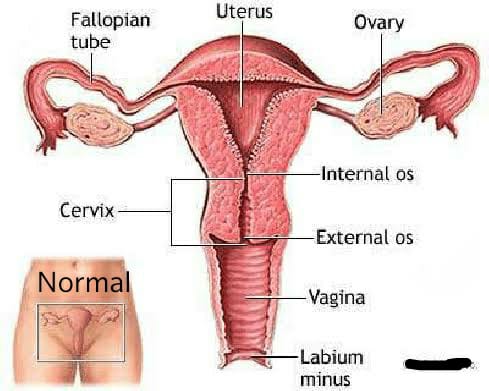
The most frequent uterine anomalies (Fig. 3) are those resulting from varying degrees of failure of fusion of the müllerian ducts. This variability makes classification difficult and determination of the true incidence uncertain. Many of these malformations are detected by radiologic or sonographic studies. Estimates of incidence vary from 0.13% to 4.0%. The incidence of müllerian anomalies in patients with infertility has been reported to be as high as 6.3%.Pregnancy occurs in many women despite these anomalies. The complication rates with pregnancy are considerably increased; complications include abortion, prematurity, postpartum hemorrhage, retained placenta, and breech presentation. Not surprisingly, the rate of cesarean delivery is markedly higher.

Uterus duplex, or the bicornuate uterus, is the most frequent uterine anomaly. The unicollis type in which there is a single cervix with a septum that does not reach the cervix is the most frequent type, occurring in over one third of all patients with uterine anomalies.

Uterus duplex bicollis, in which two cervices are present, is less frequent. Obstetric complications are frequent, but live births do occur.

Uterus didelphys, with completely separate uterine cavities, is also frequent. The cervices are externally united and the uterine fundi are externally separate. In most patients the vagina is septate, causing a double vagina. The halves of such a uterus are often of different sizes. If there is an asymmetric vaginal septum which occludes one vagina, mucocolpos or hematocolpos may result. Communicating uteri, involving an incomplete uterine septum with part of the fetus in each uterine cavity, occasionally occur.

Uterus septus is an essentially normal uterus with a septum reaching to the cervix.

Uterus subseptus involves a partial septum that does not reach the cervix. Twins apparently occur approximately three times more often in women with this condition than in women with normal uteri; the cause is not clear.
Uterus arcuatus is a normal uterus without a septum. The fundus, however, is notched or flattened. There is usually no interference with normal pregnancy.
Uterus unicornis is a uterus with a single horn. A normal vagina and a single normal tube are usually present. The other half of the uterus is usually absent or rudimentary. In most patients the kidney is missing on the side of the missing uterus. Successful pregnancy can occur.

Separate hemiuteri with separate vaginas is a rare condition that is usually associated with duplications of urethra and bladder or of the colon and anus. Pregnancy in each of the two hemiuteri in the same woman at different times has been reported.

These anomalies result from failure of fusion of the paired müllerian ducts, but in some instances there is a true duplication of the ducts on one or both sides. Such duplications result from splitting of the müllerian duct during the seventh week of development. Accessory tubes or ovaries may be present.

Semmens extensively reviewed the literature on genital tract anomalies and evaluated 56 personal patients as well as 500 cases from the literature. He employed a simplified classification, based entirely on the functional capacity of the uterine cavity, which divided genital tract anomalies into two groups: group I—hemiuterus of single müllerian origin, paired or otherwise, and group II—uterus of dual müllerian origin, associated with varying degrees of absorption. He concluded that if the entire functional component has been derived from a single müllerian duct and its vaginal outlet is a cervical canal of similar origin its capacity is smaller than that of the uterus resulting from fusion of bilateral ducts.

By contrast, if the uterus is derived from two müllerian ducts, its capacity is larger. Semmens concluded that the space available for the developing fetus as well as the variations in uterine circulation had a direct effect on the length of gestation, onset and behavior of labor, and overall fetal salvage. The diagnosis of uterine anomalies during pregnancy is always difficult. The most important factor is awareness of their existence and frequency and of the problems that frequently develop. Findings such as a floating head at term without apparent cause, notching and broadening of the uterine fundus, abnormal lie, recurrent breech presentations, prolonged third stage of labor, and trapped or retained placenta indicate the possibility of an anomaly. In abnormal uteri, triangular spasm and cornual pocketing of the placenta may occur and necessitate manual removal of the placenta.

The administration of oxytocin under these circumstances usually increases the degree of pocketing in an abnormal uterus. Among antepartum patients, pyelitis (often associated with a urologic anomaly), passage of a decidual cast, hemorrhage, and premature rupture of the membranes are the most common complications. Intrapartum complications include septal dystocia, incarcerated nongravid horn, uterine inertia, and retained placenta. Fetal dystocia is considerably increased. Postpartum complications are few and include hemorrhage, retained placenta, and failure of uterine involution. Most complications of uterine anomalies in the pregnant woman can be anticipated and managed successfully if the clinician is alert to the possibility of anomalies and if the nature of the anomaly is known.

The finding of an asymmetrically located cervix in the vaginal fornix, an excessively large cervix whether or not a septum is present, or a duplicated cervix suggests uterine abnormality. Abnormal configurations of the fundus of the uterus in the third trimester should suggest abnormalities to the clinician, as should abnormal presentations and failure of the presenting part to engage without apparent reason. An obstetric history of successive abortions, recurrent breech presentations, or recurrent pyelitis should alert the obstetrician to the possibility of anomalies and, in nonpregnant women, to the need for hysterosalpingogram, saline sonography, magnetic resonance imaging, and renal imaging.

Dr. Ruchi Tandon is a reputed Gynecologist practicing in leading hospitals in South Delhi namely Max and Apollo hospitals with over 14 years of experience in handling all kinds of Gynecological conditions including uterine abnormalities.

Diagnostic Laparoscopy for infertility

Laparoscopy overview

• Laparoscopy for infertility is a minimally invasive surgical procedure in which a telescope-like instrument (laparoscope) with light and small camera allows the surgeon to examine the pelvic anatomy for causes of female infertility.
• Laparoscopy can be diagnostic, assessing for causes of infertility in the uterus, fallopian tubes and ovaries.
• An operative laparoscopy involves surgical treatment for problems found during a diagnostic laparoscopy procedure, utilizing small surgical tools passed through the laparoscope.
• Laparoscopic surgery can also be used to remove scar tissue or endometriosis.

When is laparoscopy used for treating infertility?

Laparoscopy for infertility is generally only performed after other fertility tests have not resulted in a conclusive diagnosis. For this reason, laparoscopy is often performed on women with unexplained infertility.

Laparoscopy also allows for biopsy of suspect growths and cysts that may be hampering fertility. Laparoscopy may be recommended for women experiencing pelvic pain, which is a potential symptom of endometriosis. Laparoscopy can also remove scar tissue that can be a cause of pelvic or abdominal pain.

Why It’s Needed

Some causes of infertility can only be diagnosed through laparoscopy.

(Endometriosis, for example.) Laparoscopy allows your doctor to not only see what’s inside your abdomen but also biopsy suspicious growths or cysts.

Also, laparoscopic surgery can treat some causes of infertility, allowing you a better chance at getting pregnant either naturally or with fertility treatments.

How Is It Done?

Laparoscopy is performed in a hospital under general anesthesia. While it is sometimes possible to conduct a diagnostic laparoscopy in a fertility clinic office, this is not recommended. In the office setting, if something is found during the procedure, you will need to have the procedure again in a hospital setting for the repair.

Your doctor will give you instructions on how to prepare for surgery beforehand. You will probably be told not to eat or drink for 8 or more hours before your scheduled surgery, and you may be instructed to take antibiotics.

When you get to the hospital, you’ll receive an IV, through which fluids and medication to help you relax will be delivered. The anesthesiologist will place a mask over your face, and after breathing a sweet-smelling gas for a few minutes, you’ll fall asleep.

Once the anesthesia has taken effect, the doctor will make a small cut around your belly button. Through this cut, a needle will be used to fill your abdomen with carbon dioxide gas. This provides room for your doctor to see the organs and move the surgical instruments.

Once your abdomen is filled with gas, the surgeon will then place the laparoscope through the cut to look around at your pelvic organs. The surgeon may also biopsy tissue for testing.

The surgeon will visually evaluate the pelvic organs and the surrounding abdominal organs. He or she will look for the presence of cysts, fibroids, scar tissue or adhesions, and endometrial growths. He or she will also look at the shape, color, and size of the reproductive organs.

A dye may be injected through the cervix, so the surgeon can evaluate if the fallopian tubes are open.

Even if no signs of endometriosis or other problems are found, the surgeon may remove a sample of tissue to be tested. Sometimes, very mild endometriosis is microscopic and cannot be seen by the naked eye with the laparoscopic camera.

If an ectopic pregnancy is suspected, the surgeon will evaluate the fallopian tubes for abnormal pregnancy.

How Will It Feel?

During laparoscopic surgery, you’ll be under the effects of general anesthesia, so you should not feel any pain, nor remember the procedure.

When you wake up, you may have a sore throat. This is caused by the tube placed down your throat to help you breathe during surgery. (This tube is removed before you wake-up).

It’s normal for the area around the cuts to feel sore, and your abdomen may feel tender, especially if your doctor removes a lot of scar tissue. You may feel bloated from the carbon dioxide gas, and you may experience sharp pains in your shoulder. This should go away in a few days.

Dr. Ruchi Tandon is a reputed Gynecologist practicing in leading hospitals in South Delhi namely Max and Apollo hospitals with over 14 years of experience in handling all kinds of Gynecological conditions including infertility.

Blocked fallopian tubes

Blocked fallopian tubes are one possible cause of female infertility. There are usually no symptoms, but there are some risk factors that can increase the chance of developing the condition. The medical term for a blocked fallopian tube is tubal occlusion.

The fallopian tubes are muscular tubes that are lined with delicate hair-like structures. These “hairs” work in both directions; helping an egg to travel from the ovaries down to the womb (uterus) and helping sperm travel up from the womb.

Each fallopian tube ends in fimbriae, which are finger-like structures. The fimbriae catch and guide an egg when the ovary releases it.

The fallopian tubes play an important role in conception because they are where most eggs are fertilized.

If any part of the fallopian tube is damaged, for example by surgery or an infection, they can become blocked by scar tissue.

Symptoms

Blocked fallopian tubes do not often present symptoms other than difficulty conceiving. Doctors typically class this as having tried to conceive for 1 year without success.

A blocked fallopian tube may cause some women to experience symptoms such as pain in the pelvis or belly. This pain might happen regularly, such as around the time of their period, or be constant.

Sometimes, a blockage in a fallopian tube can cause a fertilized egg to get stuck. This is known as an ectopic pregnancy.

An ectopic pregnancy may not always cause symptoms and is usually detected during a scan. However, some woman may experience signs of pregnancy, such as stomach pain on one side of the body, or vaginal bleeding. Any woman who suspects she has an ectopic pregnancy should seek immediate medical attention.

Causes

Fallopian tubes can become blocked for a range of reasons, which include:
• a history of pelvic infection
• a previous burst appendix
• having had a sexually transmitted disease, such as gonorrhea or chlamydia
• endometriosis, a condition that causes the lining of the womb to grow outside of the uterus
• history of abdominal surgery
• hydrosalpinx, which is swelling and fluid at the end of a fallopian tube Effects on fertility

All of these conditions can affect the fallopian tubes directly or this area of the body. In most cases, these conditions or procedures create scar tissue that can block the tubes.

The female reproductive system is made up of the ovaries, uterus, and fallopian tubes.

If a medical problem has affected any of these three areas, it may make getting pregnant more difficult.

Each of the two ovaries is connected to the uterus by a fallopian tube. The ovaries store eggs and release them randomly, with one ovary releasing an egg each month.

For example, the right ovary might release an egg for 3 months in a row, and then the left ovary might release an egg the following month.

If one fallopian tube is blocked, it may still be possible for an egg to be fertilized. If both are blocked, this is less likely.

Diagnosis

Blocked fallopian tubes can be difficult to identify. The tubes can open and close, so it is not always easy to tell if they are blocked or just closed.

There are three key tests to diagnose blocked fallopian tubes:

• An X-ray test, known as a hysterosalpingogram or HSG. A doctor injects a harmless dye into the womb, which should flow into the fallopian tubes. The stain is visible on an X-ray. If the fluid does not flow into the fallopian tubes, they may have a blockage.
• An ultrasound test, known as a sonohysterogram. This is very similar to the HSG test but uses sound waves to build up a picture of the fallopian tubes.
• Keyhole surgery, known as a laparoscopy. A surgeon makes a small cut in the body and inserts a tiny camera to take pictures of the fallopian tubes from inside.

A laparoscopy is the most accurate test for blocked tubes. However, doctors may not recommend this test as an early diagnosis because it is invasive and cannot treat the issue.

A doctor may be able to suggest a possible diagnosis based on medical history. For example, a woman may have had a burst appendix in the past. If the woman has had difficulty conceiving, this could suggest blocked fallopian tubes as a likely cause.

Dr. Ruchi Tandon is a reputed Gynecologist practicing in leading hospitals in South Delhi namely Max and Apollo hospitals with over 14 years of experience in handling all kinds of Gynecological conditions including high risk Pregnancies and blocked fallopian tubes.