In a world where reproductive rights are at the forefront of social and political discussions, understanding safe abortion is crucial. Safe abortion is not just a medical procedure; it’s a complex intersection of healthcare, legislation, and societal attitudes.
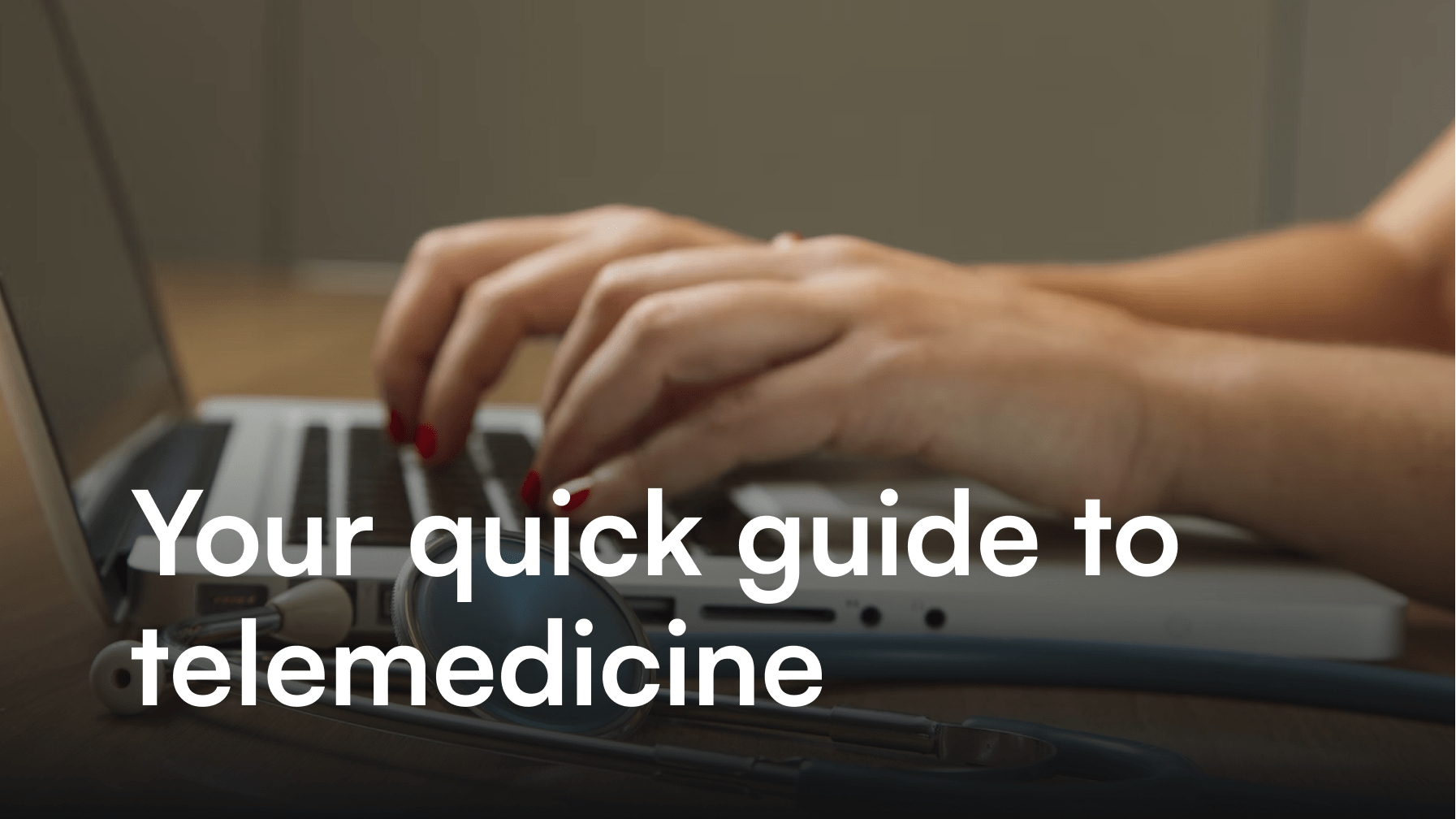
It is always carried out in the best abortion clinic in South Delhi. In this quick guide, we’ll explore key aspects of safe abortion, shedding light on the medical, legal, and ethical dimensions of this critical reproductive choice.
The Importance of Accessible Healthcare: Ensuring Safe Abortion Services
Access to safe abortion services is a fundamental aspect of reproductive rights. Lack of accessibility can lead individuals to seek unsafe alternatives, jeopardizing their health and well-being. Advocacy for accessible healthcare is not merely a call for medical services but a demand for autonomy over one’s body and reproductive choices.
Legal Landscape: Navigating Abortion Laws
Abortion laws vary globally, and staying informed about the legal landscape is essential. Some regions embrace progressive policies, guaranteeing the right to safe abortion, while others impose restrictive measures. Understanding local laws is vital for individuals and healthcare providers alike, ensuring compliance and promoting reproductive rights within the confines of legal frameworks. The Best Gynecologist in Greater Kailash can guide you with this better.
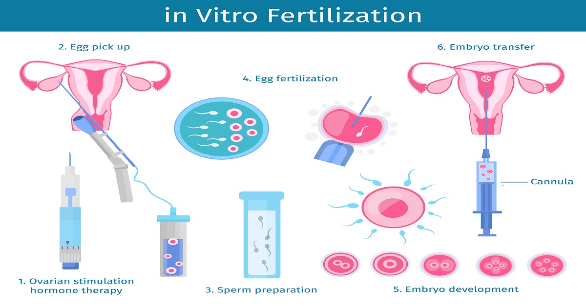
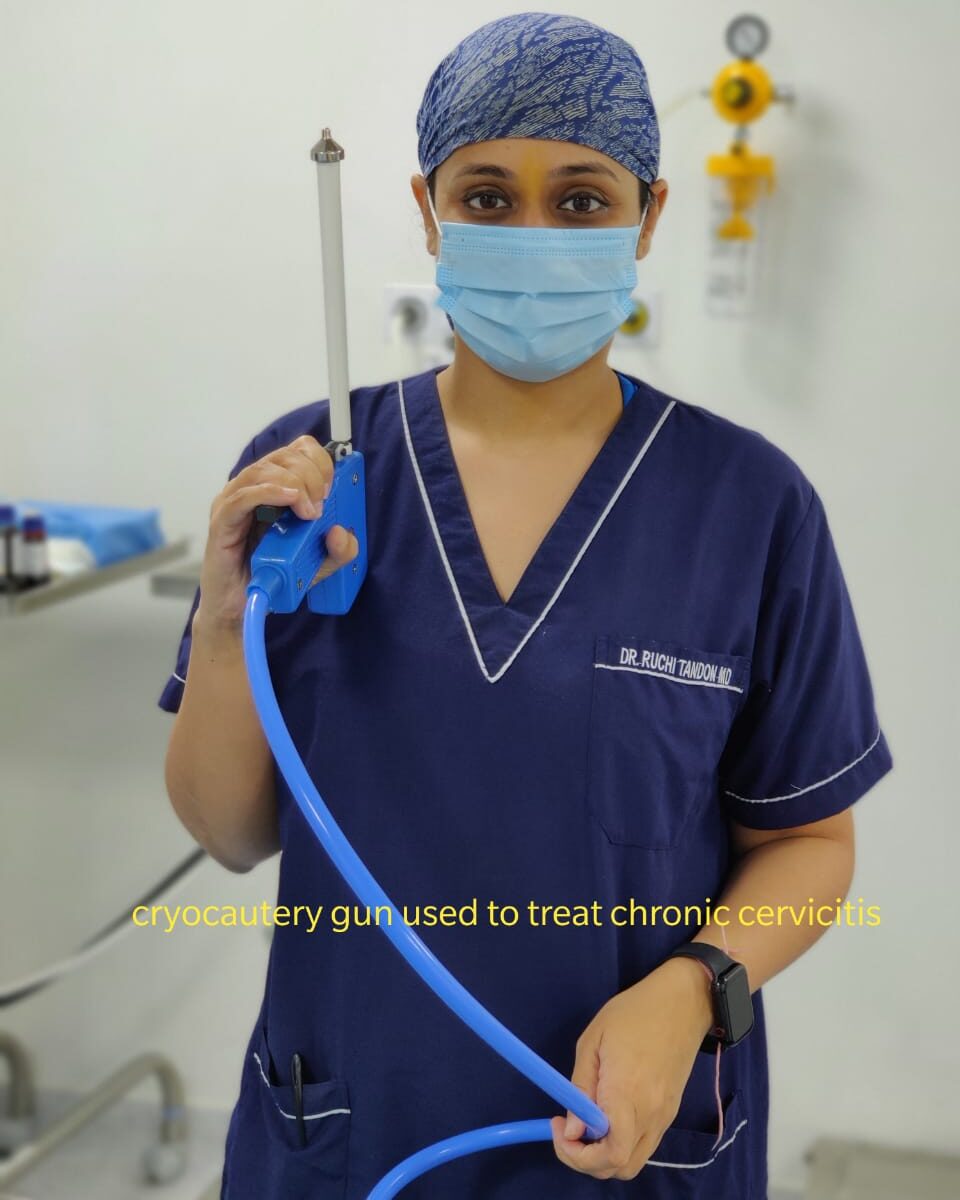
Medical Advances: Exploring Safe Abortion Procedures
Advancements in medical technology have transformed abortion procedures, making them safer and more accessible. From medication abortions to minimally invasive surgical techniques, individuals now have a range of options tailored to their specific needs. Knowledge about these procedures empowers individuals to make informed choices about their reproductive health.
Destigmatizing Abortion: Fostering Supportive Societal Attitudes
One of the significant challenges in the realm of safe abortion is the persistent stigma associated with it. Destigmatizing abortion involves fostering open conversations, challenging misconceptions, and promoting empathy. Creating a supportive societal environment is crucial for those seeking safe abortion services or the best abortion clinic in South Delhi, ensuring they are met with understanding rather than judgment.
Mental Health Considerations: Navigating Emotional Well-being
The decision to undergo an abortion can evoke a range of emotions, and prioritizing mental health is paramount. Healthcare providers play a crucial role in offering comprehensive support, including counseling services. Acknowledging the emotional aspects of abortion is integral to holistic healthcare, ensuring that individuals feel heard and supported throughout the process.
Conclusion
Safe abortion is not just a medical procedure; it’s a facet of reproductive freedom that intersects with legal, societal, and emotional dimensions. Empowering individuals with information, fostering supportive environments, and advocating for accessible healthcare are essential steps toward ensuring that safe abortion remains a fundamental right for everyone.
In the journey towards reproductive justice, knowledge is power, and compassion is key. To know more, consult Dr. Ruchi Tandon, the best gynecologist in Greater Kailash.